Diagnosis
Malignant melanoma
Malignancy derived from the malignant transformation of melanocytes.
Etiology
Cause [pmp]
UVB and UVA radiation exposure
Spontaneous mutation
Pathophysiology [pmp]
Ionizing radiation from cumulative solar damage
Atmospheric absorption
Cloud cover
Ozone layer depletion
DNA injury
Mutation in the tumor suppressor genes (BRAF)
Malignant transformation (nevus --> melanoma)
5 different histologic malignant melanomas
Superficial spreading: 70% of melanomas, any body surface
Nodular: 10-15% of melanomas, any body surface
Lentigo maligna: 10-15% of melanomas, sun-exposed areas
Acral lentiginous: palms, soles, and subungual areas
Mucosal lentiginous: 3% of the melanomas, mucosal epithelium that lines the respiratory, gastrointestinal, genitourinary tracts, conjunctiva, oral cavity and esophagus
Risk factors [pmp]
Family history
Lifetime sun exposure
Personal characteristics
Blue eyes
Fair or red hair
Pale complexion
History of sunburns
Freckled skin
More than 50 nevi
Sporadic dysplastic nevi
Congenital nevus
Immunosuppression
Complications [pmp]
Metastasis
Lymphedema after sentinel surgery
Epidemiology
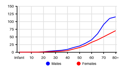
Incidence per 100.000 [mas][3eq][rsu][rtz][7nb]
Symptoms & findings
Symptoms
Clinical findings
None listed.
Anamneses
None listed.
Localized findings
Approach
Dematoscopy:
Asymmetry
Border/Bleeding: irregular, ragged, notched, or blurred edges
Color: very dark or variable
Diameter > 6 mm
Evolving: changes in size, shape, or color
Blood test: Complete blood count, ALP, ASAT, ALAT, albumin, lactate dehydrogenase
Biopsy
Sentinel node biopsy
Blood test: ALP, transaminases, albumin and LD
Chest x-ray
MR caput
CT thorax/abdomen
PET-CT
Clark staging
I - All tumor cells above basement membrane (in situ)
II - Tumor extends into papillary dermis
III - Tumor extends to interface between papillary and reticular dermis
IV - Tumor extends between bundles of collagen of reticular dermis
V - Tumor invasion of subcutaneous tissue
Breslow classification (thickness)
< 0.75 mm
0.76-1.5 mm
1.51-4 mm
> 4 mm
Treatment
Prophylaxis: Sun-protective behaviors can reduce harmful exposure to UV [ts8]
Sunscreen
Sun-protective clothing
Early-stage melanoma: Stage I-II [pmp]
Excision:
Melanoma in situ (Tis): 0.5 to 1 cm margin
T1 (≤1 mm): 1 cm margin
T2 (≥1 to 2 mm): 1 to 2 cm margin
T3 or T4 (>2 mm): 2 cm margin
Sentinel lymph node biopsy: if increased risk of regional node metastasis
Depth of melanoma (Breslow's depth) T1b or higher staging
Preoperative (neoadjuvant) therapy may be appropriate in select cases
Advanced, metastatic, unresectable, or recurrent melanoma: Adjuvant therapy
Immunotherapy: Nivolumab, pembrolizumab, ipilimumab, and relatlimab
Signal-transduction inhibitor: BRAF and MEK inhibitors
Intralesional therapy (talimogene laherparepvec)
Chemotherapy
Palliative local therapy
Stereotactic radiosurgery
External-beam radiation
Differential diagnoses
Actinic keratosis, Atypical fibroxanthoma, Basal cell carcinoma, Cutaneous T-cell lymphoma, Hemangioma, Histiocytoid hemangioma, Lentigo, Mycosis fungoides, Nevus, Sebaceous carcinoma, Squamous cell carcinoma
References
[1] Heistein JB, Acharya U, Mukkamalla SKR. Malignant Melanoma. [Updated 2024 Feb 17]. In: StatPearls [Internet]. Treasure Island (FL): StatPearls Publishing; 2025 Jan-. Available from: https://www.ncbi.nlm.nih.gov/books/NBK470409/
[2] Memon A, Bannister P, Rogers I, Sundin J, Al-Ayadhy B, James PW, McNally RJQ. Changing epidemiology and age-specific incidence of cutaneous malignant melanoma in England: An analysis of the national cancer registration data by age, gender and anatomical site, 1981-2018. Lancet Reg Health Eur. 2021 Jan 6;2:100024.
[3] Diffey BL, Frank SA. Age-specific acceleration in malignant melanoma. F1000Res. 2017 Jan 9;6:27
[4] Sneyd MJ, Cox B. Melanoma in Maori, Asian, and Pacific peoples in New Zealand. Cancer Epidemiol Biomarkers Prev. 2009 Jun;18(6):1706-13.
[5] Guy GP Jr, Thomas CC, Thompson T, Watson M, Massetti GM, Richardson LC; Centers for Disease Control and Prevention (CDC). Vital signs: melanoma incidence and mortality trends and projections - United States, 1982-2030. MMWR Morb Mortal Wkly Rep. 2015 Jun 5;64(21):591-6.
[6] Reinhart, Jacob P. et al. Incidence and mortality trends of primary cutaneous melanoma: A 50-year Rochester Epidemiologic Project study. JAAD International, Volume 16, 144 - 154
[7] http://emedicine.medscape.com/article/280245; [Medscape]