Diagnosis
Photosensitivity
An inflammatory skin disease caused by the combination of a chemical and light exposure [yhp][z2q][oep]
Phototoxic: common, large amount of agent required, may involve unexposed areas
Photoallergic: rare, small amount of agent required, strictly sun-exposed
Photosensitive epilepsy: A “photoparoxysmal” response to photic stimulation [p1p]
Etiology
Cause [z2q]
Primary (autoimmune) photodermatoses:
Polymorphic light eruption, Juvenile spring eruption, Actinic folliculitis, Actinic prurigo, Solar urticaria, Chronic actinic/photosensitivity dermatitis, Hydroa vacciniforme
Exogenous (drug/chemical-induced) photodermatosis: thiazides, tetracyclines, NSAIDs, phenothiazines, voriconazole, quinine, vemurafenib, psoralen (plants, vegetables, fruit), fragrances (cosmetics), sunscreen chemicals, dyes and disinfectants
Photo-exacerbated dermatoses: Cutaneous lupus erythematosus, lupus erythematosus, Sjogren syndrome, Darier disease, Rosacea, Melasma, Pemphigus vulgaris, Pemphigus foliaceus, Atopic dermatitis, Seborrhoeic dermatitis, Psoriasis, Lichen planus (actinicus), Erythema multiforme, Mycosis fungoides
Metabolic photodermatosis (rare): Porphyria cutanea tarda, Erythropoietic protoporphyria, Variegate porphyria, Erythropoietic porphyria (Gunther disease)
Genetic photodermatosis (very rare): Xeroderma pigmentosum, Cockayne syndrome, Trichothiodystrophy, Bloom syndrome, Rothmund Thomson syndrome
Pathophysiology
Phototoxic: common, direct damage to tissue caused by a photoactivated compound
Pathophysiology of photoactivation:
UV-A (320-400 nm) -> excitation of electrons -> excited-state electrons return to a more stable configuration -> release of energy -> formation of reactive oxygen intermediates -> damage cell membranes and DNA -> inflammatory response -> exaggerated sunburn reaction
Phototoxic agents:
Antibiotics (Tetracyclines, Fluoroquinolones, Sulfonamides)
NSAIDs (also photoallergic)
Statins (also photoallergic)
Antifungals (also photoallergic)
Amiodarone
Neuroleptics
Diuretics
Retinoid
Sulfonylurea
Porphyrin
Coal
Drugs
Psoralen
Photoallergic: rare, cell-mediated immune responses in which the antigen is a light-activated drug
Pathophysiology of photoallergy:
Antigen-presenting Langerhans cells migrate to regional lymph nodes and present photoallergen -> T cells become activated, proliferate and migrate to the site of photoallergen deposition -> T cells orchestrate an inflammatory response -> eczematous morphology
Photoallergic agents:
Sulfonylurea
Dapsone
Oral contraceptives
Sunscreens
Fragrances
Epidemiology
Incidence per 100.000
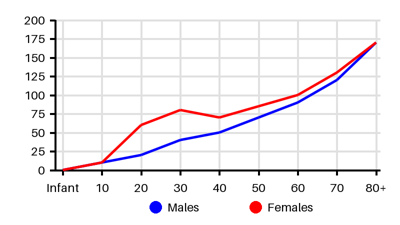
Symptoms & findings
Symptoms
Clinical findings
None listed.
Anamneses
None listed.
Localized findings
Approach
Blood: Full blood count
Urine porphyrin
Antibody levels (ANA, anti-SSA/Ro, ENA)
Photopatch
Provocation phototesting with UV
Biopsy
Treatment
Sun protection: Avoid direct sunlight, covering clothing, sunscreen
Treatment of the underlying disorder
Differential diagnoses
Acute intermittent porphyria, Dermatomyositis, Drug eruptions, Eczema, Epidermolysis bullosa, Epilepsy, Lichen planus, Systemic lupus erythematosus, Urticaria
References
[1] http://emedicine.medscape.com/article/1049648; [Medscape]
[2] Oakley AM, Badri T, Harris BW. Photosensitivity. Updated 2023 Aug 8: https://www.ncbi.nlm.nih.gov/books/NBK431072/
[3] Inamadar AC, Palit A. Photosensitivity in children: An approach to diagnosis and management. Indian J Dermatol Venereol Leprol 2005;71:73-9.
[4] Verrotti A, Beccaria F, Fiori F, Montagnini A, Capovilla G. Photosensitivity: epidemiology, genetics, clinical manifestations, assessment, and management. Epileptic Disord. 2012 Dec;14(4):349-62.
[5] de Bittencourt PR. Photosensitivity: the magnitude of the problem. Epilepsia. 2004;45 Suppl 1:30-4.
[6] https://www.uptodate.com/contents/photosensitivity-disorders-photodermatoses-clinical-manifestations-diagnosis-and-treatment
[7] Burfield L, Rutter KJ, Thompson B, Marjanovic EJ, Neale RE, Rhodes LE. Systematic review of the prevalence and incidence of the photodermatoses with meta-analysis of the prevalence of polymorphic light eruption. J Eur Acad Dermatol Venereol. 2023 Mar;37(3):511-520.