Diagnosis
Pityriasis rosea
An acute self-limiting papulosquamous disorder causing scaling rash mainly on the trunk and extremities lasting 2-10 weeks.
Also known as: Herpes tonsurans maculosus, Pityriasis circinata, Roseola annulata
Etiology
Cause [rtb]
Unknown
Infection (human herpes virus 6/7, influenza, diphtheria, smallpox, hepatitis B, pneumococcus, and COVID-19, bacteria, and spirochetes) is suspected due to:
Prodrome phase: Sore throat, gastrointestinal disturbance, fever, and arthralgia
Seasonal variation
Community clustering
Low rate of recurrence, which suggests immunity
Noninfective causes like atopy, autoimmunity, vaccination, drugs (gold compounds, captopril, barbiturates, D-penicillamine, clonidine, ACE inhibitors, NSAIDs, hydrochlorothiazide, atypical antipsychotics, imatinib, metronidazole, isotretinoin, and clozapine)
Pathophysiology [rtb]
Not entirely understood
Trigger --> T-cell–mediated immune response --> characteristic skin pattern: [2s6]
A herald ("mother") patch surrounded by scaly oval plaques on the trunk and proximal extremities forming a characteristic "Christmas tree" appearance
Complications [rtb]
Superimposed bacterial infection
Itching, discomfort, and impaired quality of life
Hyperpigmentation
Recurrence
Epidemiology
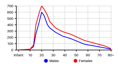
Prevalence per 100.000 [g6n][zbd][rtb][4so][bsq][woz][pws][tzm][8sn][ktu]
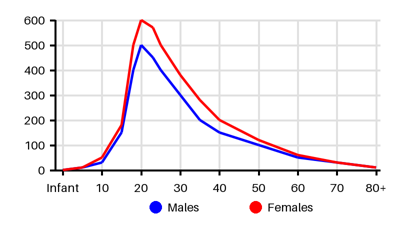
Incidence per 100.000 [g6n][zbd][rtb][4so][bsq][woz][pws][tzm][8sn][ktu]
Symptoms & findings
Symptoms
None listed.
Clinical findings
None listed.
Anamneses
None listed.
Localized findings
Approach
Dermatoscopy
Biopsy
Exclude syphilis
Diagnostic criteria [eir][yp7]
Essential clinical features:
Discrete circular or oval lesions
Scaling on most lesions, and
Peripheral collarette scaling with central clearance on at least two lesions
Optional clinical features:
Truncal and proximal limb distribution
Orientation of most lesions along skin cleavage lines
A herald patch (not necessarily the largest) appearing at least two days before eruption of other lesions
Exclusional clinical features:
Multiple small vesicles at the centre of two or more lesions
Two or more lesions on palmar or plantar skin surfaces
Clinical or serological evidence of secondary syphilis
Treatment
Self-limiting disease
Supportive therapy: [rtb][eir]
Moisturizers
Bathing with soap alternatives
Cautious sunlight exposure without burning
Emollients
Antihistamines
Therapy
Topical steroids
Acyclovir (considered in severe cases)
Macrolides (no proven benefit)
Narrowband ultraviolet B therapy
Differential diagnoses
Drug eruptions, Eczema, Erythema multiforme, Kaposi sarcoma, Lichen planus, Parapsoriasis, Pityriasis alba, Pityriasis versicolor, Psoriasis, Seborrheic dermatitis, Syphilis, Tinea corporis, Tinea versicolor
References
[1] Litchman G, Nair PA, Syed HA, et al. Pityriasis Rosea. Updated 2024 Mar 1: https://www.ncbi.nlm.nih.gov/books/NBK448091/
[2] Leung AKC, Lam JM, Leong KF, Hon KL. Pityriasis Rosea: An Updated Review. Curr Pediatr Rev. 2021;17(3):201-211.
[3] Chuh AA, Dofitas BL, Comisel GG, Reveiz L, Sharma V, Garner SE, Chu F. Interventions for pityriasis rosea. Cochrane Database Syst Rev. 2007 Apr 18;(2):CD005068.
[4] Chuang TY, Ilstrup DM, Perry HO, Kurland LT. Pityriasis rosea in Rochester, Minnesota, 1969 to 1978. J Am Acad Dermatol. 1982 Jul;7(1):80-9.
[5] Tay YK, Goh CL. One-year review of pityriasis rosea at the National Skin Centre, Singapore. Ann Acad Med Singap. 1999 Nov;28(6):829-31.
[6] Ayanlowo O, Akinkugbe A, Olumide Y. The pityriasis rosea calendar: a 7 year review of seasonal variation, age and sex distributi
[7] Harman M, Aytekin S, Akdeniz S, Inalöz HS. An epidemiological study of pityriasis rosea in the Eastern Anatolia. Eur J Epidemiol. 1998 Jul;14(5):495-7.
[8] Cheong WK, Wong KS. An epidemiological study of pityriasis rosea in Middle Road Hospital. Singapore Med J. 1989 Feb;30(1):60-2.
[9] Ganguly S. A clinicoepidemiological study of pityriasis rosea in South India. Skinmed. 2013 May-Jun;11(3):141-6.
[10] Abercrombie GF. Pityriasis rosea 1964-65. J R Coll Gen Pract. 1968 Oct;16(4):268-74.
[11] Joshi TP, Calderara GA, Lipoff JB. Prevalence of pityriasis rosea in the United States: A cross-sectional study using the All of Us database. JAAD Int. 2022 May 19;8:45-46.
[12] Eisman S, Sinclair R. Pityriasis rosea. BMJ. 2015 Oct 29;351:h5233.
[13] Chuh AA. Diagnostic criteria for pityriasis rosea: a prospective case control study for assessment of validity. J Eur Acad Dermatol Venereol. 2003 Jan;17(1):101-3.
[14] [Clinical Dermatology 4.th Edition by Weller, Hunter, Savin and Dahl]