Diagnosis
Pityriasis versicolor
A benign superficial chronic fungal infection usually characterized by hypopigmented or hyperpigmented macules and patches on the chest and the back.
Also known as: Tinea versicolor
Etiology
Cause [a83]
Malassezia furfur, formerly known as Pityrosporum
Causes infection in some individuals while remains as normal flora in others
Genetic predisposition and a hereditary component may play a role
Pathophysiology [a83]
Malassezia is a fungal inhabitant of the normal skin flora present on healthy skin (oily regions: face, scalp, and back)
Not contagious
Unknown host trigger --> Malassezia transforms into its pathogenic filamentous form --> can cause tinea versicolor
Risk factors [a83]
Oily skin: Poor hygiene, humidity, hyperhidrosis (heavy sweating), oily lotions
Immunosuppression: Steroid therapy, Cushing disease, Diabetes
Antibiotic therapy
Malnutrition
Epidemiology
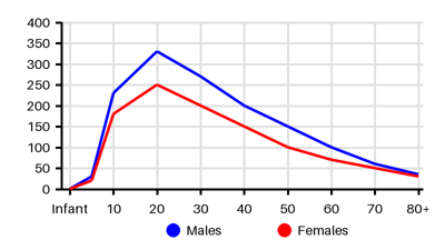
Incidence per 100.000 [a83][arn][vpi][ef7][dys]
Symptoms & findings
Symptoms
None listed.
Clinical findings
None listed.
Anamneses
None listed.
Localized findings
Approach
Rash:
Type: hyperpigmented or hypopigmented, finely scaling patches or plaques
Pattern: face (children), scalp, trunk, neck, and proximal extremities
Dermoscopy
Wood's lamp
Microscopy with KOH
Biopsy
Treatment
Topical treatment: [a83][arn]
Ketoconazole 2% shampoo for 3 days
Selenium sulfide 2.5% shampoo or lotion applied for 10 minutes daily for 1 week
Terbinafine 1% once or twice daily from 1 to 4 weeks
Ciclopirox 1% applied twice daily for 2 weeks
Zinc pyrithione 1% daily for 5 minutes for 2 weeks
Ultraviolet UV-B phototherapy (limited evidence)
Oral therapy: if resistant to topical treatment or with widespread disease
Itraconazole 200 mg daily for 7 days
Fluconazole 300 mg weekly for 2 weeks
Prophylaxis:
Topical selenium sulfide 2.5% or ketoconazole 2% shampoo applied to the entire body for ten minutes once per month
Oral Itraconazole 200 mg twice daily, once each month
Differential diagnoses
Candidiasis, Eczema, Erythrasma, Folliculitis, Mycosis fungoides, Pityriasis alba, Pityriasis rosea, Psoriasis, Seborrheic dermatitis, Syphilis, Tinea, Tinea corporis, Vitiligo
References
[1] Karray M, McKinney WP. Tinea Versicolor. Updated 2024 Feb 12: https://www.ncbi.nlm.nih.gov/books/NBK482500/
[2] http://emedicine.medscape.com/article/1091575; [Medscape]
[3] Mahmoudabadi A Z, Mossavi Z, Zarrin M. Pityriasis versicolor in Ahvaz, Iran. Jundishapur J Microbiol. 2009;2(3): 92-96.
[4] Borelli D, Jacobs PH, Nall L. Tinea versicolor: epidemiologic, clinical, and therapeutic aspects. J Am Acad Dermatol. 1991 Aug;25(2 Pt 1):300-5.
[5] He SM, Du WD, Yang S, Zhou SM, Li W, Wang J, Xiao FL, Xu SX, Zhang XJ. The genetic epidemiology of tinea versicolor in China. Mycoses. 2008 Jan;51(1):55-62.