Diagnosis
Tracheoesophageal malformation
Failure of complete separation of trachea from esophagus. Can occur with or without esophageal atresia and usually involves a tracheoesophageal fistula.
Also known as: Esophageal atresia, Tracheoesophageal fistula
Etiology
Cause/Pathophysiology [zri][goo]
The esophagus is a muscular tube that transports a food bolus from the pharynx to the stomach
Esophagus and trachea originate from a single diverticulum and divide at fifth week of gestation
The anomaly arises from a developmental disruption with failure of separation of the embryogenic foregut into trachea and esophagus during the fifth to sixth week of embryonic life
Clinical presentation:
Polyhydramnios
Drooling
Dysphagia
Respiratory tract infections
Esophagitis
Complications [goo]
Esophageal anastomotic leak after surgery
Esophageal anastomotic stricture after surgery
Esophageal dysmotility --> gastroesophageal reflux
Tracheomalacia after surgery
Classification of tracheoesophageal malformations [hui][utf][tzu][ept]
Isolated esophageal atresia without tracheoesophageal fistula (5-13%)
EA with proximal TEF and blind distal esophageal pouch (0.4-5.7%)
EA with distal TEF and blind proximal esophageal pouch (78-92%)
EA with proximal and distal TEF (<0.1-2.6%)
Isolated TEF (2.4-6.5%)
Associated syndromes and anomalies:
Down's syndrome, Edward's syndrome, Feingold syndrome, CHARGE (Coloboma, Heart, Choanal, Retarded growth, Genital, Ear), VACTERL (Vertebral, Anorectal, Cardiac, TracheoEsophageal fistula, Renal and Limb), Potter sequence
Cardiovascular anomalies in 23% (most common)
Ventricular or atrial septal defects
Patent ductus arteriosus
Tetralogy of Fallot
Right-sided aortic arch
Persistence of left superior vena cava
Musculoskeletal malformations
Radial limb abnormalities
Polydactyly
Lower limb defects
Hemivertebrae
Rib abnormalities
Scoliosis
Anorectal and intestinal malformations
Pyloric stenosis
Duodenal atresia
Imperforate anus
Omphalocele
Malrotation
Genitourinary malformations
Hypospadias
Renal agenesis
Ureteral and urethral abnormalities
Horseshoe kidney
Polycystic kidney disease
Head and neck anomalies
Microcephaly
Micrognathia
Other craniofacial anomalies
Pulmonary and mediastinal anomalies
Tracheobronchomalacia
Pulmonary hypoplasia
Tracheal stenosis or atresia
Tracheal upper pouch
Less common defects may include
Single umbilical artery
Diaphragmatic hernia
Nervous system abnormalities, such as cerebral hypoplasia or spina bifida
Environmental factors:
Higher paternal and maternal age
Low parity
Maternal diabetes mellitus or maternal phenylketonuria
Frequent maternal hot tub use
In utero exposure to diethylstilbestrol, methimazole, carbimazole, or mycophenolate
Epidemiology
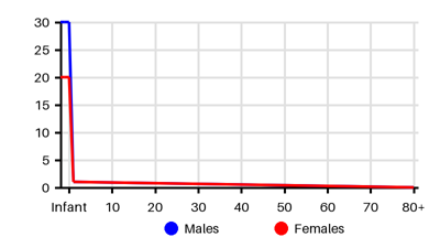
Incidence per 100.000 [yv9][g9z][zri][u1e][goo][hui][tzu]
Symptoms & findings
Symptoms
Apnea, Choking, Drooling, Intercostal retraction, Respiratory distress, Salivation, Small for gestational age
Clinical findings
Anamneses
Approach
Impossibility of the passage of an orogastric catheter beyond 11 or 12 centimeters [tzu]
Prenatal ultrasound
Chest x-ray
Abdominal x-ray
MRI
Treatment
Survival is only possible with surgical correction.
Neonatal intensive care: [tzu]
Ventilatory: endotracheal tube distal to the fistula
Nutritional support
Antibiotics
Damage control: [tzu]
Gastrostomy
Estimation of the gap between the proximal and distal esophagus
Lengthening of the esophagus
Delayed surgical intervention at three months of age: [tzu]
Right posterolateral extrapleural thoracotomy
Closure of the fistula: divided near the trachea and sewn
Correction of the EA:
Upper esophageal pouch is identified with downward tension on an oro-esophageal tube
Placement of a traction suture at the end of the pouch
Dissection to mobilize the proximal pouch to the level of the thoracic inlet
Esophageal continuity is accomplished with an end-to-end anastomosis
Optional trans-anastomotic tube
Long-gap EA (>3 cm) presents a challenge:
Colonic interposition
Gastric tube interposition
Gastric transposition
Free jejunal graft interposition
Differential diagnoses
Esophageal diverticula, Esophageal perforation, Esophageal rupture, Esophageal stricture, Gastroesophageal reflux disease, Pneumonia, Pneumonitis
References
[1] Pedersen RN, Calzolari E, Husby S, Garne E; EUROCAT Working group. Oesophageal atresia: prevalence, prenatal diagnosis and associated anomalies in 23 European regions. Arch Dis Child. 2012 Mar;97(3):227-32.
[2] Baldwin DL, Yadav D. Esophageal Atresia. Updated 2023 Jul 25: https://www.ncbi.nlm.nih.gov/books/NBK560848/
[3] Brosens E, Ploeg M, van Bever Y. Clinical and etiological heterogeneity in patients with tracheo-esophageal malformations and associated anomalies. Eur J Med Genet. 2014 Aug;57(8):440-52.
[4] Scott, DA. Esophageal Atresia/Tracheoesophageal Fistula Overview. GeneReviews 2014 June 12: https://www.ncbi.nlm.nih.gov/books/NBK5192/
[5] Pinheiro PF, Simoes e Silva AC, Pereira RM. Current knowledge on esophageal atresia. World J Gastroenterol. 2012 Jul 28;18(28):3662-72.
[6] Broemling N, Campbell F. Anesthetic management of congenital tracheoesophageal fistula. Paediatr Anaesth. 2011 Nov;21(11):1092-9, commentary can be found in Paediatr Anaesth 2012 Mar;22(3):301.
[7] Centers for Disease Control and Prevention (CDC). Improved national prevalence estimates for 18 selected major birth defects--United States, 1999-2001. MMWR Morb Mortal Wkly Rep. 2006 Jan 6;54(51):1301-5.
[8] Schieppati A, Henter JI, Daina E, Aperia A. Why rare diseases are an important medical and social issue. Lancet. 2008 Jun 14;371(9629):2039-41.
[9] Salik I, Paul M. Tracheoesophageal Fistula. Updated 2024 Jul 2: https://www.ncbi.nlm.nih.gov/books/NBK535376/