Diagnosis
Esophageal perforation
Trauma to esophagus that may result in life-threatening leakage of air and esophageal contents into mediastinum.
Also known as: Boerhaave syndrome, Esophageal rupture
Etiology
Cause [oqu][qtv]
Iatrogenic: Endoscopy, dilatation, tamponade tubes, resection, radiotherapy
Vomiting (Boerhaave syndrome)
Mallory-Weiss syndrome
Foreign body ingestion
Esophageal ulcer
Eosinophilic esophagitis
Trauma
Esophageal cancer
Ingestion of corrosive substances
Pathophysiology [qtv]
The esophagus is anatomically divided into 3 regions based on blood syupply:
The cervical esophagus
The thoracic esophagus
The abdominal esophagus
Esophageal perforations can arise in any of the 3 natural constrictions:
Cervical: The cricopharyngeal sphincter at the level of the C6 vertebra
Thoracic: Where the esophagus is crossed by the aortic arch and left bronchus
Diaphragmatic: Where the esophagus pierces the diaphragm at the T10 vertebra
Intraluminal injuries:
Instrumentation
Ingestion of foreign bodies
Esophagitis and carcinoma
Infection or barotrauma
Extraluminal injuries
Traumatic injuries
Surgical accidents/Iatrogenic injuries: fundoplication or esophageal myotomy
Perforation --> leak of contents (gastric contents, saliva, biliary fluid):
Chemical mediastinitis
Mediastinal emphysema
Bacterial translocation
Exudative pleural effusion
Sepsis/shock
Complications [oqu][qtv]
Sepsis
Mediastinitis
Intrathoracic abscess
Respiratory failure
Shock
90-day mortality: 22-39% [cko]
Epidemiology
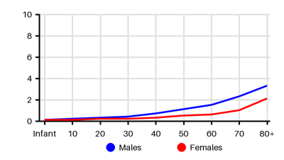
Incidence per 100.000 [cko][ds7][c8f][pyq][wmn]
Symptoms & findings
Symptoms
Crepitations, Cyanosis, Dysphagia, Dysphonia, Dyspnea, Fever, Hamman's sign, Hematemesis, Hypotension, Melena, Odynophagia, Subcutaneous emphysema, Tachycardia, Tachypnea, Vomiting
Clinical findings
Contrast extravasation, Emphysema, Mediastinal widening, Peritonitis, Pleural effusion, Pneumomediastinum, Pneumoperitoneum, Pneumothorax
Anamneses
None listed.
Localized findings
Approach
Chest x-ray
Contrast esophagography/Barium enema
Gastroscopy
CT thorax
Thoracentesis (low pH, elevated amylase)
Treatment
Initial therapy: [fon][qtv] Conservative if absence of infection and contained perforation
Admission to ICU: Hemodynamic monitoring, volume resuscitation, and stabilizatio
Nasogastric suction
Parenteral nutrition
Intravenous broad-spectrum antibiotics and antifungals
Intravenous proton pump inhibitors
Percutaneous drainage of any fluid collections
Feeding jejunostomy tube
Analgesic
Endoscopic: [cko][qou][qtv]
Stent + Thoracic drain
Endoluminal negative-pressure therapy
Endoscopic fibrin sealant
Endoscopic suture ligation
Open surgery: [fon][cko][oqu]
Primary repair ± local flap (transthoracic or transhiatal) + Thoracic drain
Esophageal resection + pericardial/pleural flap
Cervical esophagostomy:
Diversion prevents further contamination
High morbidity of the procedure
Reserved for unstable patient, major disruption of the esophageal wall, severe contamination
Differential diagnoses
Achalasia, Acute pancreatitis, Aortic dissection, Esophageal cancer, Esophageal diverticula, Esophageal spasm, Gastroesophageal reflux disease, Mallory-Weiss tear, Myocardial infarction, Perforated ulcer, Pericarditis, Pleural empyema, Pneumonia, Pneumonitis, Pneumothorax, Pulmonary embolism
References
[1] https://emedicine.medscape.com/article/425410
[2] Kassem MM, Wallen JM. Esophageal Perforation and Tears. [Updated 2024 Aug 19]. In: StatPearls [Internet]. Treasure Island (FL): StatPearls Publishing; 2025 Jan-. Available from: https://www.ncbi.nlm.nih.gov/books/NBK532298/
[3] Edholm D, Andersson RE, Frankel A. Esophageal perforations - a population-based nationwide study in Sweden with survival analysis. Scand J Gastroenterol. 2022 Sep;57(9):1018-1023.
[4] Søreide JA, Konradsson A, Sandvik OM, Øvrebø K, Viste A. Esophageal perforation: clinical patterns and outcomes from a patient cohort of Western Norway. Dig Surg. 2012;29(6):494-502.
[5] Vidarsdottir H, Blondal S, Alfredsson H, Geirsson A, Gudbjartsson T: Oesophageal perforations in Iceland: a whole population study on incidence, aetiology and surgical outcome. Thorac Cardiovasc Surg 2010;58:476–480.
[6] Ryom P, Ravn JB, Penninga L, et al: Aetiology, treatment and mortality after oesophageal perforation in Denmark. Dan Med Bull 2011;58:A4267.
[7] Khan, J., Laurikka, J., Laukkarinen, J., Toikkanen, V., & Ukkonen, M. (2020). The incidence and long-term outcomes of esophageal perforations in Finland between 1996 and 2017 – a national registry-based analysis of 1106 esophageal perforations showing high early and late mortality rates and better outcomes in patients treated at high-volume centers. Scandinavian Journal of Gastroenterology, 55(4), 395–401.
[8] Hasimoto CN, Cataneo C, Eldib R, Thomazi R, Pereira RS, Minossi JG, Cataneo AJ. Efficacy of surgical versus conservative treatment in esophageal perforation: a systematic review of case series studies. Acta Cir Bras. 2013 Apr;28(4):266-71.
[9] Carrott PW Jr, Low DE. Advances in the management of esophageal perforation. Thorac Surg Clin. 2011 Nov;21(4):541-55.
[10] Mubang RN, Sigmon DF, Stawicki SP. Esophageal Trauma. [Updated 2023 Jul 25]. In: StatPearls [Internet]. Treasure Island (FL): StatPearls Publishing; 2025 Jan-. Available from: https://www.ncbi.nlm.nih.gov/books/NBK470161/