Diagnosis → Ophthalmology
Scleritis
Inflammation of the sclera with or without adjacent corneal/uveal involvement:
Subtypes:
Anterior or posterior
Diffuse, nodular, or necrotizing
Also known as: Logaditis, Scleral inflammation, Sclerotitis
Etiology
Cause [fup][zth]
Idiopathic
Infectious (4-10%)
Virus: Herpes zoster/HSV
Bacteria: Syphilis, tuberculosis, Lyme disease
Fungal: Post-trauma, surgery, immunocompromised
Parasite
Autoimmune disease (50%):
Rheumatoid arthritis
Systemic lupus erythematosus
Ankylosing spondylitis
Reactive arthritis
Psoriatic arthritis
Gouty arthritis
Inflammatory bowel diseases
Relapsing polychondritis
Polymyositis
Sjögren syndrome
Mixed connective tissue disease
Progressive systemic sclerosis
Polyarteritis nodosa
Allergic angiitis of Churg-Strauss syndrome
Granulomatosis with polyangiitis (Wegener granulomatosis)
Behçet disease
Giant cell arteritis
Cogan syndrome
Malignancy
Surgery (pterygium removal and scleral buckle procedures)
Trauma
Medications: Pamidronate, alendronate, risedronate, zoledronic acid, ibandronate
Miscellaneous (2%): Atopy, rosacea, foreign bodies, chemical injuries
Pathophysiology [fup]
Trigger
Systemic autoimmune disease
Local ocular trigger (surgery/trauma/infection)
Scleral immune activation
Inflammatory cell infiltration and cytokine-mediated tissue injury
In necrotizing forms: vasculitis of deep episcleral/scleral vessels
Structural damage and extension
Scleral edema → severe ocular pain and deep injection
Scleral thinning/necrosis
Spillover to cornea/uvea → keratitis/uveitis, secondary glaucoma
Vision loss from keratitis, uveitis, glaucoma, cataract, retinal/choroidal complications
Necrotizing and posterior scleritis cases pose a higher risk of visual loss
Complications [zth][kvi]
Keratopathy: Corneal thinning, keratitis, punctate keratopathy --> Decreased vision
Iridocyclitis
Glaucoma
Cataract
Fundus abnormalities: Choroidal folds, subretinal mass, disk edema, macular edema, annular ciliochoroidal detachment, or serous retinal detachment
Epidemiology
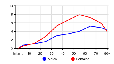
Incidence per 100.000 [fup][qun][kvi][sr2]
Default
Symptoms & findings
Symptoms
Blurred vision, Lacrimation, Photophobia, Red eye
Clinical findings
None listed.
Anamneses
None listed.
Localized findings
Approach
Slit lamp light
Red-free light
Differentiate from superficial episcleritis:
Upon installation of 2.5% topical phenylephrine in the affected eye, the blood vessels will blanch with episcleritis but will not blanch with scleritis
Blood tests: CRP, CBC, ESR, Rheumatoid factor, ANA, ANCA, HLA-B27, IgE, uric acid, HBsAg, Quantiferon, angiotensin-converting enzyme, lysozyme
Serology: Syphilis and Lyme disease
Anergy skin test: Sarcoidosis
Prick test: Atopy
Radiology:
B-scan
Sinus films
CT thorax
CT orbita
MR orbita
Scleral or corneoscleral biopsy:
Necrotizing granulomatous scleritis
Diffuse/nodular nongranulomatous scleritis
PCR of orbital abscess drainage, aqueous humor, and vitreous
Treatment
Associated disease-specific treatment [zth]
Rosacea
Gout
Atopy
Infection: Topical (and systemic) antimicrobial therapy
Noninfectious scleritis:
Topical therapy:
Topical corticosteroid eyedrops
Periorbital and subconjunctival steroid injection (often insufficient)
Systemic treatment:
1st line: Systemic NSAIDs
Indomethacin 50 mg x 3 per day
Ibuprofen 600 mg x 3 per day
Naproxen 500 mg x 2 per day
2nd line: Corticosteroids
Prednisone 1 mg/kg/day (60-80 mg/day)
Dose continued until one month after the resolution of scleritis
3rd line: Immunomodulatory drugs:
Methotrexate 7.5-25 mg PO/IM once a week
Azathioprine 1-2.5 mg/kg/day PO
Cyclophosphamide 1.5-3 mg/kg/day PO
Mycophenolate mofetil 1 g PO bid
Infliximab 3-5 mg/kg IV q6-8wk
Adalimumab 40 mg every 2 weeks
Surgery may rarely be required to preserve the integrity of the globe in cases of pending perforation
Scleral grafts from fresh donor sclera or glycerin-preserved sclera
Differential diagnoses
Conjunctivitis, Episcleritis, Foreign body, Glaucoma, Herpes zoster, Iridocyclitis, Keratitis, Periorbital cellulitis, Rosacea, Squamous cell carcinoma, Subconjunctival hemorrhage
References
[1] Lagina A, Ramphul K. Scleritis. [Updated 2023 Jun 26]. In: StatPearls [Internet]. Treasure Island (FL): StatPearls Publishing; 2026 Jan-. Available from: https://www.ncbi.nlm.nih.gov/books/NBK499944/
[2] https://emedicine.medscape.com/article/1228324 (6th september 2026)
[3] Xu TT, Reynolds MM, Hodge DO, Smith WM. Epidemiology and Clinical Characteristics of Episcleritis and Scleritis in Olmsted County, Minnesota. Am J Ophthalmol. 2020 Sep;217:317-324.
[4] Homayounfar G, Nardone N, Borkar DS, Tham VM, Porco TC, Enanoria WT, Parker JV, Vinoya AC, Uchida A, Acharya NR. Incidence of scleritis and episcleritis: results from the Pacific Ocular Inflammation Study. Am J Ophthalmol. 2013 Oct;156(4):752-8.
[5] Braithwaite T, Adderley NJ, Subramanian A, Galloway J, Kempen JH, Gokhale K, Cope AP, Dick AD, Nirantharakumar K, Denniston AK. Epidemiology of Scleritis in the United Kingdom From 1997 to 2018: Population-Based Analysis of 11 Million Patients and Association Between Scleritis and Infectious and Immune-Mediated Inflammatory Disease. Arthritis Rheumatol. 2021 Jul;73(7):1267-1276.