Diagnosis → Ophthalmology
Glaucoma
A progressive optic neuropathy with or without visual field defects, often in conjunction with elevated intraocular pressure ≥ 21 mmHg.
Etiology
Causes [wpc]
Primary open-angle glaucoma (POAG): most common
Increased resistance to aqueous outflow --> Ineffective drainage system
Rise in IOP --> Reduced blood flow to the optic nerve
Slow and painless damage to the optic nerve
Characteristic damage patterns in the visual field and the optic nerve:
Loss of peripheral vision in one or typically both eyes
Eventually loss of central vision (permanent and irreversible)
Primary angle-closure glaucoma (PACG): 10% of glaucoma cases
Darkness/drugs --> pupillary dilatation --> thick iris
Age-related lens thickening --> pushes the iris anteriorly
Closure of the angle formed between the cornea and the iris
Blockage of the outflow tract --> IOP rapidly increases
Blurry vision
Corneal edema
Eye pain
Headache
Nausea
Emesis
Secondary glaucoma arises due to obstruction/damage to trabecular meshwork
Lens subluxation in Marfan syndrome
Irregular corneal endothelium migration onto the trabecular meshwork
Neovascularization (retinal artery/vein occlusion, proliferative diabetic retinopathy)
Peripheral anterior synechiae
Eye injury
Post-ophthalmic surgery (ciliary body edema)
Sulfa drugs --> ciliochoroidal effusion
Pseudoexfoliation (flaky material within the anterior chamber that clog the trabecular meshwork)
Pigment dispersion syndrome (pigment granules from the posterior iris surface)
Steroid-induced glaucoma
Carotid-cavernous fistula
Ellingson syndrome (uveitis-glaucoma-hyphema syndrome)
Glaucomatocyclitic crisis (Posner-Schlossman syndrome)
Inflammatory glaucoma: Uveitis/iridocyclitis
Normal-tension glaucoma
Optic nerve damage due to normal pressure, atherosclerosis or vascular insufficiency
Associated with migraines, Raynaud phenomenon, autoimmune diseases, ischemic vascular diseases, and coagulopathies
Congenital/infantile glaucoma
Affects individuals between the ages of 1 and 36 months
Trabeculodysgenesis
Anterior segment dysgenesis
Pathophysiology [wpc]
Aqueous humor is produced by the non-pigmented epithelial cells and is crucial in maintaining intraocular pressure and nourishing ocular tissues
Continuous drainage: the trabecular meshwork --> Schlemm canal --> episcleral veins
A decline in aqueous humor drainage --> elevated IntraOcular Pressure (IOP)
Glaucoma has strong correlation with IOP ≥ 21 mmHg
Risk factors [wpc]
IOP ≥ 21mmHg
Older age
Hypermetropia (shallow anterior chamber)
An angle between the iris and the cornea ≤ 20° during gonioscopy
Anterior chamber depth of less than 2.5 mm
Complications
Visual field loss
Blindness
Epidemiology
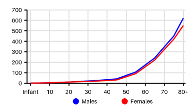
Incidence per 100.000 [bie][pon]
Default
Symptoms & findings
Symptoms
Blindness, Blurred vision, Headache, Nausea, Red eye, Scotoma, Visual disturbances, Vomiting
Clinical findings
None listed.
Anamneses
None listed.
Localized findings
Approach
Blood tests: CBC count, ESR
Serelogy for syphilis
Serum protein electrophoresis (autoimmune etiology)
Clinical exxamination:
Slit lamp examination of the anterior segment
Fundoscopic examination
Visual field testing
Tonometry (Goldmann applanation tonometry)
Gonioscopy
Pachymetry
Optical coherence tomography (OCT)
Confocal scanning laser ophthalmoscopy
Scanning laser polarimetry
Neuroimaging
Diagnostic criteria
Optic neuropathy: Loss of retinal ganglion cells/axons within the optic nerve
Characteristic optic nerve head appearance
Progressive loss of vision
Usually in conjunction with elevated intraocular pressure ≥ 21 mmHg
Treatment
Goal: impossible to reverse vision loss. Treatment aims to lower IOP [wpc]
Medications: [wpc][ihy]
Prostaglandin analogs
β-blockers
Carbonic anhydrase inhibitors
α-2 agonists
Miotic agents
Rho-kinase inhibitors
Nitric oxide-donating medications
Hyperosmotic agents (isosorbide dinitrate, mannitol, glycerin)
Laser:
Laser trabeculoplasty: benefits last months, and retreatments are necessary
Laser ablation of the ciliary body to reduce aqueous production
Laser peripheral iridotomy (acute angle-closure glaucoma)
Surgery:
Trabeculectomy
Deep sclerectomy
Canaloplasty
Insertion of a drainage valve/tube shunt
Minimally invasive glaucoma surgery (MIGS) [ihy]
Deep sclerectomy/viscocanalostomy
360-degree suture canaloplasty
Trabectome
Endoscopic cyclophotocoagulation (ECP)
iStent implant
Gonioscopy-assisted transluminal trabeculotomy (GAAT)
Kahook dual blade
Xen Gel Implant
Differential diagnoses
Cluster headache, Conjunctivitis, Corneal abrasion, Corneal ulcer, Endophthalmitis, Episcleritis, Hyphema, Iridocyclitis, Keratitis, Migraine, Optic neuritis, Periorbital cellulitis, Subconjunctival hemorrhage
References
[1] Dietze J, Blair K, Zeppieri M, et al. Glaucoma. [Updated 2024 Mar 16]. In: StatPearls [Internet]. Treasure Island (FL): StatPearls Publishing; 2026 Jan-. Available from: https://www.ncbi.nlm.nih.gov/books/NBK538217/
[2] Velez-Nandayapa L, Eggenschwiler L, Jick SS, Meier CR, Becker C. The incidence rates and prevalence of treated glaucoma in the United Kingdom, 2000-2022. PLoS One. 2026 Sep 3;21(9):e0354463.
[3] Shan S, Wu J, Cao J, Feng Y, Zhou J, Luo Z, Song P, Rudan I; Global Health Epidemiology Research Group (GHERG). Global incidence and risk factors for glaucoma: A systematic review and meta-analysis of prospective studies. J Glob Health. 2024 Nov 8;14:04252.
[4] https://emedicine.medscape.com/article/1206147 (12th September 2026)