Diagnosis → Ophthalmology
Keratitis
Inflammation of the cornea.
Also known as: Corneal infection, Corneal ulcer
Etiology
Cause [43s][eci]
Bacterial: Staphylococcus spp. (direct invasion or antigen-mediated), Streptococcus spp. (blocked nasolacrimal duct), Pseudomonas aeruginosa (contact lens–related), Serratia spp., Haemophilus influenzae, Moraxella spp., Enterobacteriaceae, MRSA, Nocardia spp., nontuberculous mycobacteria (post-surgical)
Fungal: Fusarium spp., Aspergillus spp., Candida spp. (post-transplant, immunocompromised); dematiaceous fungi, Curvularia spp.
Viral: HSV, VZV, adenovirus, EBV, enterovirus
Parasitic: Acanthamoeba spp., microsporidia, Onchocerca volvulus, Pythium
Non-infectious:
Trauma: Trichiasis, foreign body, Giant papillary conjunctivitis
Immune-mediated connective tissue disease: Rheumatoid arthritis, sarcoidosis, granulomatosis with polyangiitis, polyarteritis nodosa, systemic lupus erythematosus, ocular rosacea, vernal/atopic keratoconjunctivitis
Neurotrophic corneal ulcer: Herpes zoster ophthalmicus, trigeminal nerve damage
Exposure keratopathy --> Xerophthalmia
Toxic/medication-related: NSAIDs, anesthetics, preservatives, corticosteroids
Pathophysiology [43s]
Disruption of corneal epithelial barrier → loss of the primary defense mechanism
Contact lens wear, trauma, surgery, ocular surface disease, Vitamin A deficiency
Pathogen adherence and stromal invasion
Inflammation --> polymorphonuclear leukocytes and macrophages
Inflammatory cells --> Proteases/hydrolase/toxins --> Tissue destruction
Starts with epithelial ulceration: Usually seen in HSV → dendritic ulceration
Stromal infiltration: Immune-mediated disciform or necrotizing stromal keratitis
Host inflammatory response
Corneal edema
Leukocyte infiltration
Neovascularization
Anterior chamber reaction (hypopyon in bacterial/fungal)
Severe pain
Risk Factors [eci]
Contact lens wear
Ocular trauma
Previous ocular surgery
Ocular surface disease: dry eye, blepharitis, neurotrophic keratopathy, lid malposition
Topical corticosteroids
Systemic immunosuppression / HIV
Substance abuse
Developing-world with limited access to care
Complications [43s]
Corneal scarring and opacification
Corneal melting (keratolysis) --> thinning --> perforation:
Endophthalmitis
Iris prolapse
Anterior dislocation of the lens
Anterior capsular cataract
Corneal fistula
Purulent uveitis
Intraocular hemorrhage
Corneal scarring- nebula, macula, leucoma, adherent leucoma
Keratectasia
Disorganized anterior chamber
Autoevisceration
Phthisis bulbi
Secondary glaucoma
Iridocyclitis
Cataract
Recurrent disease
Permanent monocular blindness
Epidemiology
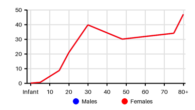
Incidence per 100.000 [oss][pnc][wtb][k1w]
Default
Symptoms & findings
Symptoms
Blindness, Blurred vision, Hypoesthesia, Keratic precipitates, Lacrimation, Mucopus, Photophobia, Red eye
Clinical findings
Anamneses
Localized findings
Approach
Slit-lamp examination: Ulcer size, location, depth, and margins of infiltrate/ulcer, fluorescein staining, anterior chamber reaction
Bacterial: suppurative infiltrate >1 mm, indistinct edges, epithelial defect, hypopyon
Fungal: feathery borders, fixed hypopyon, satellite lesions
HSV epithelial: characteristic dendritic ulcer with terminal bulbs
Acanthamoeba: ring infiltrate, perineural infiltrates, severe pain, contact lens history
Corneal scraping for smear and culture (gold standard):
Infiltrate is central and/or >2 mm with stromal involvement or melting
Chronic or unresponsive to empiric broad-spectrum antibiotics
History of corneal surgery or transplant
Multifocal infiltrates
Sight-threatening/severe presentation
Corneal smear stains:
Gram stain (bacteria; sensitivity 60–75%)
Giemsa stain (bacteria, fungi, Acanthamoeba; sensitivity 40–85% for fungi)
Calcofluor white-KOH (fungi, Acanthamoeba)
Acid-fast stain (Mycobacteria, Nocardia)
Corneal culture
PCR/NAAT
HSV/VZV NAAT (high sensitivity 70–100%)
Acanthamoeba NAAT (most sensitive)
Multiplex PCR for bacteria if culture-negative
Do not use on contact lens case/solution (high false-positive rate)
In vivo confocal microscopy: sensitivity 80–94% for fungal keratitis
Anterior segment OCT: Measure of infiltrate size, depth, and corneal thinning
Treatment
Remove contact lenses immediately [43s]
Obtain corneal scrapings for smear and culture before starting antimicrobials
Cycloplegic agent (cyclopentolate or atropine 1%)
Xerophthalmia: Vitamin A supplementation
Mild bacterial keratitis
Topical fluoroquinolone monotherapy 3–4 times/day
Ciprofloxacin 0.3%
Ofloxacin 0.3%
Levofloxacin 1.5%
MRSA: Vancomycin or topical linezolid 0.2%
Pseudomonas: Oral doxycycline is added and topical imipenem-cilastatin
Nocardia: Topical fortified amikacin (2.5%)
Atypical mycobacteria: Topical fortified amikacin (4%) or Clarithromycin (2%)
Severe/central bacterial keratitis (infiltrate >2 mm, stromal melting, hypopyon)
Complete coverage against both gram-positive and gram-negative organisms
Fortified cefazolin 5%
Tobramycin
Gentamicin
Protozoal keratitis: Biguanides and pentamidine
Oomycete keratitis: Topical linezolid (0.2%) + topical and oral azithromycin
Fungal keratitis: Albendazole (400 mg twice daily for 3-4 weeks) and topical fumagillin
Viral keratitis: Topical Acyclovir 3% x 5 (+ oral Acyclovir 400 mg x 5)
Herpes zoster ophthalmicus: Oral Acyclovir 800 mg x 5 for 1 week
Local causes --> correction of pathology:
Trichiatic lashes: electrolysis of eyelash hair root follicle
Distichiasis: lid splitting procedure
Entropion: lid everting procedure
Giant papillary conjunctivitis: Topical steroids + topical cyclosporine + lubricants
Removal of a foreign body
Systemic causes: Topical and oral steroids or methotrexate
Perforated corneal ulcer:
Cyanoacrylate tissue adhesive glue or amniotic membrane graft
Differential diagnoses
Blepharitis, Conjunctivitis, Corneal abrasion, Corneal ulcer, Endophthalmitis, Entropion, Foreign body, Glaucoma, Herpes zoster, Iridocyclitis, Keratoconjunctivitis sicca, Nasolacrimal duct obstruction, Periorbital cellulitis, Rosacea, Scleritis, Sebaceous carcinoma, Stevens-Johnson syndrome, Subconjunctival hemorrhage, Toxic epidermal necrolysis
References
[1] Singh P, Gupta A, Tripathy K. Keratitis. [Updated 2023 Aug 25]. In: StatPearls [Internet]. Treasure Island (FL): StatPearls Publishing; 2026 Jan-. Available from: https://www.ncbi.nlm.nih.gov/books/NBK559014/
[2] Ting DSJ, Ho CS, Deshmukh R, Said DG, Dua HS. Infectious keratitis: an update on epidemiology, causative microorganisms, risk factors, and antimicrobial resistance. Eye (Lond). 2021 Apr;35(4):1084-1101.
[3] Erie JC, Nevitt MP, Hodge DO, Ballard DJ. Incidence of ulcerative keratitis in a defined population from 1950 through 1988. Arch Ophthalmol. 1993 Dec;111(12):1665-71.
[4] Jeng BH, Gritz DC, Kumar AB, Holsclaw DS, Porco TC, Smith SD, Whitcher JP, Margolis TP, Wong IG. Epidemiology of ulcerative keratitis in Northern California. Arch Ophthalmol. 2010 Aug;128(8):1022-8.
[5] Ting DSJ, Ho CS, Cairns J, Elsahn A, Al-Aqaba M, Boswell T, Said DG, Dua HS. 12-year analysis of incidence, microbiological profiles and in vitro antimicrobial susceptibility of infectious keratitis: the Nottingham Infectious Keratitis Study. Br J Ophthalmol. 2021 Mar;105(3):328-333.
[6] Koh YY, Sun CC, Hsiao CH. Epidemiology and the Estimated Burden of Microbial Keratitis on the Health Care System in Taiwan: A 14-Year Population-Based Study. Am J Ophthalmol. 2020 Dec;220:152-159.
[7] Stapleton F. The epidemiology of infectious keratitis. Ocul Surf. 2023 Apr;28:351-363.