Diagnosis
Eosinophilic esophagitis
A chronic immune/antigen-mediated inflammatory disorder causing eosinophilic infiltration of the esophagus and esophageal dysfunction.
Etiology
Cause [zrk]
Unknown
Environmental: food allergy
Genetic: family history and correlation to atopy (allergy, asthma, atopic dermatitis)
Host immune factors
Patophysiology [zrk]
There are no physiologic eosinophils in the esophagus
Eosinophils present in the esophagus in response to various stimuli or antigen
Mucosal inflammation --> remodeling of esophageal mucosa --> fibrosis
Smooth muscle dysfunction --> dysphagia, epigastric/chest pain, dyspepsia, and food impaction
Disease course:
Spontaneous remission with or without treatment
Waxing and waning course
Progressive course leading to strictures
Risk factors [wfm]
Aeroallergens
Food allergens
Helicobacter pylori (protective)
Proton pump inhibitors
Cold or arid climates
Population density (urban vs rural locations)
Early life factors (antibiotic use; cesarian section)
Connective tissue disorders
Epidemiology
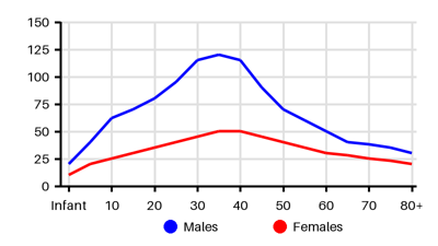
Prevalence per 100.000 [wfm][dwy][im6][siq][psb]
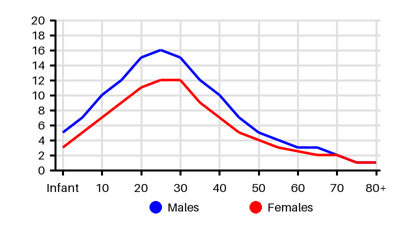
Incidence per 100.000 [wfm][dwy][im6][siq][psb]
Symptoms & findings
Symptoms
Diarrhea, Dyspepsia, Dysphagia, Failure to thrive, Feeding difficulty, Heartburn, Nausea, Vomiting
Clinical findings
Esophageal stricture, Esophageal ulcer, Gastroesophageal Reflux Disease
Anamneses
None listed.
Localized findings
Approach
Endoscopy
Biopsy
Diagnostic criteria [im6]
Esophageal dysfunction and
≥15 eosinophils/high-power field (HPF) on endoscopic biopsy
Treatment
Dietary therapy: exclusions of antigenic foods [zrk]
If unknown allergenic food or agent: a trial of food elimination diet
Common allergenic food: milk, wheat, peanut, egg, soy, and seafood/shellfish
Proton pump inhibitors 20-40 mg daily or twice daily
Topical steroids: Budesonide 1-2 mg per day
Systemic steroids: Prednisone 2 mg/kg/day (max 60mg/day)
Endoscopic dilation in cases with strictures
Immunsuppresion:
Monoclonal antibody: Dupilumab [ryh]
Differential diagnoses
Achalasia, Celiac disease, Crohn disease, Drug side effects, Eosinophilia, Eosinophilic gastroenteritis, Gastroesophageal reflux disease, Graft-versus-host disease, Mixed connective tissue disease, Pemphigus vulgaris, Vasculitis
References
[1] Roussel JM, Pandit S. Eosinophilic Esophagitis. [Updated 2023 Aug 8]. In: StatPearls [Internet]. Treasure Island (FL): StatPearls Publishing; 2025 Jan-. Available from: https://www.ncbi.nlm.nih.gov/books/NBK459297/
[2] Dellon ES. Epidemiology of eosinophilic esophagitis. Gastroenterol Clin North Am. 2014 Jun;43(2):201-18.
[3] Arias Á, Pérez-Martínez I, Tenías JM, Lucendo AJ. Systematic review with meta-analysis: the incidence and prevalence of eosinophilic oesophagitis in children and adults in population-based studies. Aliment Pharmacol Ther. 2016 Jan;43(1):3-15.
[4] Hahn JW, Lee K, Shin JI, Cho SH, Turner S, Shin JU, Yeniova AÖ, Koyanagi A, Jacob L, Smith L, Fond G, Boyer L, Lee SW, Kwon R, Kim S, Shin YH, Rhee SY, Moon JS, Ko JS, Yon DK, Papadopoulos NG. Global Incidence and Prevalence of Eosinophilic Esophagitis, 1976-2022: A Systematic Review and Meta-analysis. Clin Gastroenterol Hepatol. 2023 Dec;21(13):3270-3284.e77.
[5] Soon IS, Butzner JD, Kaplan GG, deBruyn JC. Incidence and prevalence of eosinophilic esophagitis in children. J Pediatr Gastroenterol Nutr. 2013 Jul;57(1):72-80.
[6] Dellon ES, Jensen ET, Martin CF, Shaheen NJ, Kappelman MD. Prevalence of eosinophilic esophagitis in the United States. Clin Gastroenterol Hepatol. 2014 Apr;12(4):589-96.e1.
[7] https://emedicine.medscape.com/article/1610470
[8] Furuta G.T., Katzka D.A.: Eosinophilic esophagitis. NEJM 2015; 373: pp. 1640-1648.
[9] Buckmeier, Bridget K. et al. The Incidence and Prevalence of Eosinophilic Esophagitis. The Journal of Allergy and Clinical Immunology 121 (2008).