Diagnosis
Staphylococcal scalded skin syndrome
A systemic cutaneous infection caused by toxin-producing staphylococcal species causing blistering and acute exfoliation of the skin.
Also known as: Pemphigus neonatorum, Ritter disease
Etiology
Cause [4i6]
Staphylococcus aureus (exotoxin-producing strain are less than 5%)
Pathophysiology [4i6][tsp]
Some strains of Staphylococcus aureus produce exfoliative toxins
The exfoliative toxins are spread hematogenously from a localized source
Nasopharynx
Conjunctivae
Staphylococcal pneumonia
Bacteremia
The toxins act as proteases that target the protein desmoglein-1 (a cell-to-cell attachment protein found only in the superficial epidermis)
Loss of cellular adhesion among keratinocytes
Severety varies:
A few blisters localized to the site
Severe exfoliation affecting almost the entire body (dangerous)
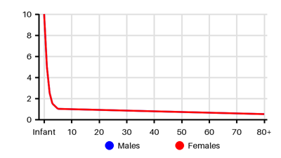
SSSS is much less common in adults because of neutralizing antibodies and efficient renal clearance of exotoxins from the bloodstream
Loss of the protective skin barrier --> risk for complications
Risk factors [4i6]
Children
Immunocompromised adults
Complications [4i6]
Hypothermia
Dehydration from fluid loss
Secondary infections
Electrolyte imbalance
Sepsis
Acute kidney injury
Scarring
Epidemiology
Incidence per 100.000 [4i6][zkt][tsp][svn][q1a]
Symptoms & findings
Symptoms
Edema, Fever, Irritability, Malaise, Nikolsky's sign
Clinical findings
Elevated CRP, Elevated Sedimentation Rate, Leukocytosis
Anamneses
None listed.
Localized findings
Approach
Blood test: electrolytes, renal function
PCR
Bulla culture
Culture with S. aureus from skin, umbilicus, nasopharynx, conjunctiva, and diaper area
Blood culture
Gram stain
Skin biopsy
Treatment
Fluid resuscitation (denuded skin --> water loss) [4i6]
IV antibiotics: [4i6][tsp]
Penicillinase-resistant Cefazolin 50-100 mg/kg daily, divided every 8 hours
Clindamycin
Dicloxacillin 500 mg every 6 hours
Trimethoprim/sulfamethoxazole 160/800 mg every 12 hours
Vancomycin if MRSA
Skin care:
Wound cleansing
Moist dressings/Vaseline
Antiseptic or hydrocolloid ointment and creams
Monitoring to avoid hypothermia and hemodynamic instability
Differential diagnoses
Acute exanthematic pustulosis, Burn wound, Cellulitis, Drug side effects, Eczema, Epidermolysis bullosa, Erysipelas, Erythema multiforme, Gas gangrene, Graft-versus-host disease, Impetigo, Kawasaki disease, Necrotizing fasciitis, Pemphigus foliaceus, Pemphigus vulgaris, Scarlet fever, Stevens-Johnson syndrome, Toxic epidermal necrolysis, Toxic shock syndrome
References
[1] Saleh HM, Ross A, Sathe NC. Staphylococcal Scalded Skin Syndrome. [Updated 2025 Jun 22]. In: StatPearls [Internet]. Treasure Island (FL): StatPearls Publishing; 2025 Jan-. Available from: https://www.ncbi.nlm.nih.gov/books/NBK448135/
[2] Hooper SM, Fung CM, Torr C, Lawrence SM. Case Report: Delayed recurrence of staphylococcal scalded skin syndrome in an extremely low birth weight infant. Front Pediatr. 2025 Mar 17;13:1564633.
[3] Lipový B, Brychta P, Chaloupková Z, Suchánek I. Staphylococcal scalded skin syndrome in the Czech Republic: an epidemiological study. Burns. 2012 Mar;38(2):296-300.
[4] Arnold JD, Hoek SN, Kirkorian AY. Epidemiology of staphylococcal scalded skin syndrome in the United States: A cross-sectional study, 2010-2014. J Am Acad Dermatol. 2018 Feb;78(2):404-406.
[5] Mockenhaupt M, Idzko M, Grosber M, Schöpf E, Norgauer J. Epidemiology of staphylococcal scalded skin syndrome in Germany. J Invest Dermatol. 2005 Apr;124(4):700-3.
[6] http://emedicine.medscape.com/article/788199; [Medscape]
[7] Staiman A, Hsu DY, Silverberg JI. Epidemiology of staphylococcal scalded skin syndrome in U.S. children. Br J Dermatol. 2018 Mar;178(3):704-708.