Diagnosis
Necrotizing fasciitis
A rapidly progressive life-threatening infection characterized by widespread necrosis of the subcutaneous tissue and the fascia.
Etiology
Causes [tbd]
Type I necrotizing fasciitis (80%): mixed infection (elderly patients)
Bacteroides, Clostridium, Peptostreptococcus, Enterobacteriaceae, Escherichia coli, Proteus, Pseudomonas, Klebsiella, Vibrio vulnificus, Group A beta-hemolytic streptococci, Haemophilus aphrophilus, S aureus, varicella infection and NSAIDs.
Type II necrotizing fasciitis:
Group A beta-hemolytic Streptococcus (Streptococcus pyogenes) alone or in combination with Staphylococcus aureus
Type III necrotizing fasciitis:
Vibrio or Aeromonas species (contaminated water or food)
Combination with an underlying cause:
Diabetes
Atherosclerotic vascular disease
Venous insufficiency
Pathophysiology [why]
Streptococcal pyrogenic exotoxins are directly toxic --> release of cytokines
The infection typically travels along the fascial plane, which has a poor blood supply
Vascular occlusion --> ischemia --> tissue necrosis
Superficial nerve damage --> localized anesthesia
Sepsis due to systemic toxicity
The overlying tissues are initially unaffected --> delaying diagnosis and treatment
Skin affection after several days:
Color: Erythema with purple to bluish-gray hue
Texture: Indurated
Swollen, shiny, and warm in temperature
Painful out of proportion
Bullae
Cutaneous gangrene
Risk factors
Trauma
Surgery
Insect bites
Immunocompromised patients: diabetes mellitus, cancer, alcoholism, vascular insufficiencies, organ transplants, HIV infection, or neutropenia
Complications [why]
Multiorgan failure
Septic shock
Loss of extremity
Severe scarring
Toxic shock
Death
Epidemiology
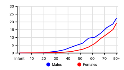
Incidence per 100.000 [tbd][myi][rba]
Symptoms & findings
Symptoms
Anesthesia, Anorexia, Diarrhea, Edema, Fever, Hypotension, Malaise, Myalgia, Pruritus, Subcutaneous emphysema, Tachycardia
Clinical findings
Elevated Blood Urea Nitrogen, Elevated Creatinine, Hyponatremia, Leukocytosis, Shock
Anamneses
Localized findings
Approach
The Laboratory Risk Indicator for Necrotizing Infection Score ≥ 6p [why]
CRP > 150 (4p)
White cell count >25 (2p) >15 (1p)
Hemoglobin <11 (2p) <13.5 (1p)
Na < 135 (2p)
Creatinine > 140 (2p)
Blood gas
Urinanalysis
Blood culture
Tissue culture
Biopsy
PCR
Ultrasound
CT
MRI
Treatment
Transferred immediately to the intensive care unit: [why]
Aggressive resuscitation with fluids and inotropes to maintain blood pressure
Antibiotics (wide-spectrum):
Imipenem 1 g every 6 to 8 hours or Piperacillin/tazobactam 4 g every 6 hours
Daptomycin 6 mg/kg QD
Clindamycin 600 mg to 900 mg 4 times daily
Meropenem 1 g IV every 8 hours
Vancomycin 15 to 20 mg/kg/dose every 8 to 12 hours
Clindamycin 600 mg to 900 mg 4 times daily.
Surgery without delay: extensive, wide debridement of all necrotic tissues
Wounds must be left open and packed with wet gauze for second look
Parenteral nutrition
Differential diagnoses
Cellulitis, Epididymitis, Erysipelas, Fournier gangrene, Gas gangrene, Inguinal hernia, Nonclostridial myonecrosis, Orchitis, Sepsis, Septic arthritis, Testicular torsion, Toxic shock syndrome
References
[1] Liu TJ, Tai HC, Chien KL, Cheng NC. Predisposing factors of necrotizing fasciitis with comparison to cellulitis in Taiwan: A nationwide population-based case-control study. J Formos Med Assoc. 2020 Jan;119(1 Pt 1):18-25
[2] Wallace HA, Perera TB. Necrotizing Fasciitis. [Updated 2023 Feb 21]: https://www.ncbi.nlm.nih.gov/books/NBK430756/
[3] Nawijn F, de Gier B, Brandwagt DAH, Groenwold RHH, Keizer J, Hietbrink F. Incidence and mortality of necrotizing fasciitis in The Netherlands: the impact of group A Streptococcus. BMC Infect Dis. 2021 Dec 6;21(1):1217
[4] Bodansky, D.M.S., Begaj, I., Evison, F. et al. A 16-year Longitudinal Cohort Study of Incidence and Bacteriology of Necrotising Fasciitis in England. World J Surg 44, 2580–2591 (2020)
[5] http://emedicine.medscape.com/article/2051157 (2014-02-10); [Medscape]
[6] http://www.uptodate.com/contents/necrotizing-soft-tissue-infections; [Uptodate]