Diagnosis
Kawasaki disease
An autoimmune vasculitis that involves small- to medium-sized arteries with mucocutaneous clinical findings such as rash, conjunctivitis, cheilitis, and glossitis.
Also known as: Mucocutaneous lymph node syndrome
Etiology
Cause [uvd][drk]
Unknown
Enviromental factors (infection): wind-borne or water-borne pathogen
Autoimmune/genetic predisposition: HLA serotypes, family history
Pathophysiology [drk][uvd]
Stage 1: The acute febrile stage [drk]
Abrupt onset of fever and mucocutaneous clinical findings.
Vasculitis: The endothelial cells and the vascular media become edematous.
Immunological activation: Neutrophils, CD8+ lymphocytes, IgA–producing plasma cells, cytokines (TNF, VEGF), interleukins, and MMPs
Vascular damage (sometimes transmural necrosis --> leading to aneurysms
Stage 2: The subacute stage [drk]
Desquamation of the digits, thrombocytosis.
The inflammatory cells are replaced by fibroblasts and monocytes
Fibrous connective tissue begins to form within the vessel wall
The vessel wall eventually becomes narrowed or occluded owing to stenosis
Vasospasm, thrombosis, rupture of aneurysm --> cardiac complications (9%)
Stage 3: The convalescent phase [drk]
Complete resolution of clinical signs of the illness, usually within 3 months
Cardiac abnormalities may still be apparent and coronary artery aneurysms may rupture in adult life
Complications [uvd]
Heart failure
Myocarditis
Valvulitis
Pericarditis
Myocardial infarction (MI)
Rupture of the coronary arteries
Ischemia-related dysrhythmias
Death
Risk of oronary artery aneurysms [uvd]
Fever >8 days (most important risk factor)
Recurrence of fever after a period of being afebrile for 48 hours
Male (3 times more likely to develop giant aneurysms)
Cardiomegaly
<1 year old of age
Lower IgG levels
Increased pro-BNP levels
Increased TNF-alpha levels
Thrombocytopenia at initial presentation
Incomplete Kawasaki Disease Diagnosis
Epidemiology
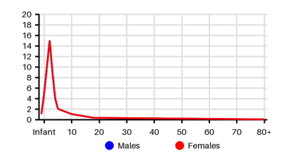
Incidence per 100.000 [iem][yg3][rnr]
Symptoms & findings
Symptoms
Anorexia, Arthralgia, Arthritis, Beau lines, Cheilitis, Conjunctivitis, Cough, Dehydration, Diarrhea, Edema, Enanthem, Facial nerve paralysis, Fever, Glossitis, Hepatomegaly, Irritability, Lethargy, Lymphadenopathy, Neck stiffness, Pyuria, Rhinorrhea, Strawberry tongue, Vomiting, Weakness
Clinical findings
Anemia, Elevated ALAT, Elevated Alpha-1-antitrypsin, Elevated Bilirubin, Elevated CRP, Elevated Sedimentation Rate, Elevated Troponin, Gallbladder wall thickening, Jaundice, Leukocytosis, Liver failure, Meatitis, Myocarditis, Orchitis, Otitis media, Pathologic Q wave, Pericardial effusion, Pericarditis, Pneumonitis, Prolonged PR interval, Renal failure, ST depression, ST elevation, Thrombocytosis, Urethritis, Uveitis, Vulvitis
Anamneses
None listed.
Localized findings
Approach
ECG
Echocardiography
MRI
MR angiography
Coronary angiography
Chest x-ray
Abdominal ultrasound
Ultrasound scrotum
Arthrocentesis
Lumbar puncture
Skin biopsy
Diagnostic criteria [yg3]
Fever of at least five days duration
Plus at least four of the following features:
Polymorphous rash
Bilateral non-purulent conjunctivitis
Changes in the peripheries
Oropharyngeal changes
Cervical lymphadenopathy
Treatment
Intravenous immunoglobulin: 2 g/kg given as a single infusion over 10–12 hours [yg3]
Failure to respond --> second dose
Failure to respond --> intravenous methylprednisolone
Aspirin: 50–100 mg/kg/day is given acutely until the fever defervesces [yg3]
Aspirin 2–5 mg/kg/day until echocardiography at six weeks after diagnosis is normal
If abnormal echocardiography --> continue Aspirin
Anti-cytokine therapies: unproven but have occasionally been used [yg3]
Differential diagnoses
Adenovirus, Cellulitis, Leptospirosis, Lyme disease, Lymphadenopathy, Mastoiditis, Measles, Meningitis, Mercury toxicity, Mononucleosis, Myocardial infarction, Parvovirus B19, Peritonsillar abscess, Polyarteritis nodosa, Retropharyngeal abscess, Rheumatic fever, Rheumatoid arthritis, Rocky mountain spotted fever, Scarlet fever, Staphylococcal scalded skin syndrome, Systemic lupus erythematosus, Toxic epidermal necrolysis, Toxic shock syndrome, Typhus
References
[1] Owens AM, Plewa MC. Kawasaki Disease. [Updated 2023 Jun 26]. In: StatPearls [Internet]. Treasure Island (FL): StatPearls Publishing; 2025 Jan-. Available from: https://www.ncbi.nlm.nih.gov/books/NBK537163/
[2] http://emedicine.medscape.com/article/965367; [Medscape]
[3] Elakabawi K, Lin J, Jiao F, Guo N, Yuan Z. Kawasaki Disease: Global Burden and Genetic Background. Cardiol Res. 2020 Feb;11(1):9-14.
[4] Burgner D, Harnden A. Kawasaki disease: What is the epidemiology telling us about the etiology? International Journal of Infectious Diseases. Volume 9, Issue 4, July 2005, Pages 185-194.
[5] Taslakian EN, Wi CI, Seol HY, Boyce TG, Johnson JN, Ryu E, King KS, Juhn YJ, Choi BS. Long-term Incidence of Kawasaki Disease in a North American Community: A Population-Based Study. Pediatr Cardiol. 2021 Jun;42(5):1033-1040.
[6] Lin MT, Wu MH. The global epidemiology of Kawasaki disease: Review and future perspectives. Glob Cardiol Sci Pract. 2017 Oct 31;2017(3):e201720.