Diagnosis
Lymphadenopathy
Enlarged cervical lymph nodes reactive to infection, inflammatory conditions, or neoplasm.
Also known as: Infectious lymphadenitis, Lymphadenitis, Reactive lymphadenitis
Etiology
Cause [ne6][dtq][nuo][wte]
Infectious
Viral:
Acute: Rhinovirus, adenovirus, influenza, parainfluenza, respiratory syncytial virus, herpes simplex virus, measles, mumps, rubella, varicella
Subacute or chronic: EBV, CMV, HIV
Bacterial:
Acute: Staphylococcus aureus, group A streptococcus, anaerobic
Subacute or chronic: Bartonella, Salmonella, Syphilis, and Yersinia
Neonates: Group B streptococcus
Zoonosis: Bartonella henselae (Cat scratch disease), Toxoplasmosis, Tularemia, Brucellosis, Histoplasmosis, Trypanosomiasis
Mycobacterium tuberculosis and atypical mycobacterial
Fungal
Parasites: toxoplasmosis, Chagas
Malignancies
Lymphoma
Leukemia
Rhabdosarcoma
Neuroblastoma
Metastasis (lung, GI, breast, kidney, nasopharyngeal and thyroid cancer)
Autoimmune disease
Sarcoidosis
Rheumatoid arthritis
Serum sickness
Systemic lupus erythematosus
Inborn metabolic storage disorder
Niemann-Pick disease
Gaucher disease
Exposure to toxic/medication
Allopurinol, atenolol, captopril, carbamazepine, cephalosporins, gold, hydralazine, penicillin, phenytoin, primidone, para methylamine, quinidine, sulfonamides, and sulindac
Congenital neck mass
Thyroglossal duct cyst
Dermoid
Branchial cleft
Lymphovascular malformation
Hemangioma
Ectopic thymus
Miscellaneous
Kikuchi-Fujimoto disease
Rosai-Dorfman disease
Langerhans cell histiocytosis
Kawasaki disease
Castleman disease
Pathophysiology [wte]
Lymphatic fluid represents the body's afferent extracellular (interstitial) fluid
Lymphatic channels conduct this fluid and label antigens with antigen-presenting cells
The lymphatic channels converge regionally to form discreet lymph nodes
The lymph node functions as an antigen filter and exposes B-cell lymphocytes, T-cell lymphocytes, and macrophages to the interstitial antigens (immune surveillance)
In case of a immune response, there is multiplication of the responding resistant cell line; thus, the node increases in size
Cell lines can commonly multiply by 3 to 5 times in 6 to 24 hours
Epidemiology
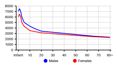
Incidence per 100.000 [fnd][sbh][b4c][ne6]
Symptoms & findings
Symptoms
Clinical findings
None listed.
Anamneses
None listed.
Localized findings
Approach
Lymph node assessment: [wte]
Age:
Neonates: congenital infection (CMV or Toxoplasma gondii)
3-5 years old: Upper respiratory tract infections, pharyngitis, otitis, or conjunctivitis
Adolescents: Sexually transmitted disease
Duration:
Acute enlargement --> acute viral or bacterial infection
Chronic lymphadenopathy lasts more than 4–6 weeks --> medications (phenytoin, isoniazid, pyrimethamine), malignancy, immunologic disorders, or Kawasaki disease
Location: May reflects the lymphatic drainage of their location
Unilateral suggest bacterial infections
Bilateral suggest viral infections (influenza, adenovirus)
Generalized: EBV, CMV, HIV, lymphoma
Anatomic: Submandibular, submental, jugular, cervical, pectoral, suboccipital, postauricular, supraclavicular, axillar, epitrochlear, inguinal
Size: Pathological size vary depending on age and location (rough estimate > 1cm)
Overlying skin changes (discolouration, induration)
Pain: Tender (infection) or non-tender (rule out malignancy)
Consistency: soft (infection/inflammation), rubber (may suggest malignancy)
Clustered may indicate malignancy
Fixed or mobile
Surface: smooth or irregular
Recent infections: tooth, mouth ulcer, cough, impetigo, cellulitis, sexually transmitted disease
History of foreign travel
Exposure to animals or insect bites
Consumption of under-cooked food/untreated water
Recent immunizations
Medications
Red flags: Weight loss, night sweats, long-lasting fever, easy bruising, joint pain/swelling, rash
Blood test: blood cell count, WBC, CRP, sedimentation rate, liver enzymes, lactate dehydrogenase, uric acid
Monospot: Streptococcus pyogenes antigens
Induced sputum
Serelogy: EBV, HIV, CMV, Parvovirus B 19, Bartonella spp., Brucella spp., Toxoplasma gondii, tuberculosis (IGRA), syphilis, Herpes simplex virus, Hepatitis B
Blood smear
Chest x-ray: mediastinal widening or lymph node due to lymphoma and sarcoidosis
Ultrasonography: size and monitoring
CT collum
Fine-needle aspiration
Biopsy - The gold standard for diagnosis of lymphadenopathy
CT abdomen
MRI
Treatment
Treat the underlying condition [wte]
Treatment of the neoplasm
Bacterial disease: Supportive care, antibiotics, and elimination of nidus of infection
Viral disease: Observation and supportive care or treatment of the virus if possible
Toxin or medication exposure: Removal of offending medication/toxin if possible
Differential diagnoses
Acute sinusitis, Branchial cleft cyst, Breast cancer, Brucellosis, Cat scratch disease, Cellulitis, Coxsackievirus infection, Cytomegalovirus, Dental infection, Dermoid Cyst, Drug side effects, Fungal infection, Hemangioma, Herpes simplex, Histoplasmosis, HIV, Influenza, Kawasaki disease, Leukemia, Lymphoma, Malignant melanoma, Mononucleosis, Neuroblastoma, Parotitis, Pharyngitis, Rhabdomyosarcoma, Rheumatoid arthritis, Sarcoidosis, Sjogren syndrome, Squamous cell carcinoma, Syphilis, Systemic lupus erythematosus, Thyroglossal duct cyst, Thyroid cancer, Toxoplasmosis, Tuberculosis, Tularemia, Typhoid fever
References
[1] Weinstock MS, Patel NA, Smith LP. Pediatric Cervical Lymphadenopathy. Pediatr Rev. 2018 Sep;39(9):433-443.
[2] Deosthali A, Donches K, DelVecchio M, Aronoff S. Etiologies of Pediatric Cervical Lymphadenopathy: A Systematic Review of 2687 Subjects. Glob Pediatr Health. 2019 Jul 27;6:2333794X19865440.
[3] Rosenberg TL, Nolder AR. Pediatric cervical lymphadenopathy. Otolaryngol Clin North Am. 2014 Oct;47(5):721-31.
[4] Freeman AM, Matto P. Lymphadenopathy. [Updated 2023 Feb 20]. In: StatPearls [Internet]. Treasure Island (FL): StatPearls Publishing; 2025 Jan-. Available from: https://www.ncbi.nlm.nih.gov/books/NBK513250/
[5] Pecora F, Abate L, Scavone S, Petrucci I, Costa F, Caminiti C, Argentiero A, Esposito S. Management of Infectious Lymphadenitis in Children. Children (Basel). 2021 Sep 27;8(10):860.
[6] Herzog LW. Prevalence of lymphadenopathy of the head and neck in infants and children. Clin Pediatr. 1983 Jul;22(7):485-7.
[7] Larsson LO, Bentzon MW, Berg Kelly K, Mellander L, Skoogh BE, Strannegård IL, Lind A. Palpable lymph nodes of the neck in Swedish schoolchildren. Acta Paediatr. 1994 Oct;83(10):1091-4.