Diagnosis
Osteosarcoma
The most common malignant bone tumor originating from primitive bone-forming mesenchymal cells.
Etiology
Cause [g5i]
Primary osteosarcoma originates within normal bone (childhood and adolescence):
Unknown cause
Possible theories:
Genetic predisposition
Enviromental factors
Secondary osteosarcoma originates in bone affected by a disease:
Paget disease of bone
Radiation exposure
Case reports: history of electrical burns, trauma, and joint arthroplasty
Pathophysiology [g5i]
Localization:
Primary: Near the growth plates of long bones (femur, tibia, humerus)
Rapid bone growth during adolescence --> Increased risk of DNA mutation
Secondary: Skull, jaw, and pelvis, primarily the ilium
Inflammation --> Increased risk of DNA mutation
Sporadic mutation in the tumor suppressor genes
Regulatory mechanisms become defective
Uncontrolled cellular proliferation of a single clone of bone cells
Osteosarcoma usually starts in osteoblasts
Clinical presentation: bone pain, swelling, pathological fracture
Risk factors [g5i]
Past treatment with chemotherapy or radiation therapy
RB1 gene
Bloom syndrome
Diamond-Blackfan anemia
Li-Fraumeni syndrome
Paget disease
Hereditary retinoblastoma
Rothmund-Thomson syndrome
Werner syndrome
Complications [g5i]
Metastasis (10-20% present with metastases, primarily to the lungs)
Pathologic fracture
Treatment complications
Classification of bone tumors
Malignant: Osteosarcoma, Chondrosarcoma, Ewings Tumor
Benign: Osteoid Osteoma, Osteoblastoma, Osteochondroma, Enchondroma, Giant cell tumor
Epidemiology
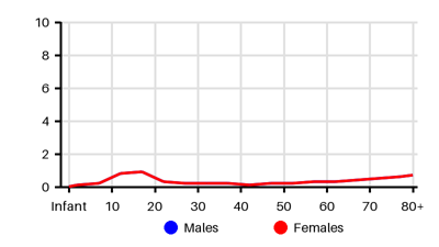
Incidence per 100.000 [g5i][sk6][gk6][d7u]
Symptoms & findings
Symptoms
Arthralgia, Cough, Fever, Hemoptysis, Lymphadenopathy, Malaise, Night sweats
Clinical findings
Anemia, Bone lesion, Codmans triangle, Elevated CRP, Elevated Sedimentation Rate, Leukocytosis, Lytic bone lesion, Onion skin, Pathologic fracture, Permeative bone lesion, Sclerotic bone lesion, Sunray appearance
Anamneses
None listed.
Localized findings
Approach
Blood test: blood count, alkaline phosphatase, and lactate dehydrogenase
X-ray
MRI
Biopsy
CT scan of the lungs
PET-CT scan
Treatment
Chemotherapy: [gk6]
Neoadjuvant (preoperative) --> Tumor shrinkage --> Facilitate surgical removal
Provide an important risk parameter
Radiotherapy: Osteosarcomas are not particularly responsive to radiotherapy
Surgery is the only option for definitive tumor removal [g5i]
Wide local excision
Limb-sparing surgery with allograft bone replacement or metallic prosthetics
Amputation
Rotationplasty
Samarium: A radioactive drug that targets areas where bone cells are growing
Targeted therapy: Drugs that block the action of specific enzymes, proteins, or other molecules involved in the growth of benign or malignant tumor cells
Kinase inhibitor
Mammalian target of rapamycin (mTOR) inhibitor [ree]
Differential diagnoses
Arthritis, Ewing sarcoma, Fracture, Langerhans cell histiocytosis, Lymphoma, Osteoblastoma, Osteoid osteoma, Osteomyelitis, Osteoperiostitis, Spinal disc herniation
References
[1] Greenwood AC, Arora RD, Shaikh H. Osteosarcoma (Osteogenic Sarcoma) [Updated 2024 Dec 11]. In: StatPearls [Internet]. Treasure Island (FL): StatPearls Publishing; 2025 Jan-. Available from: https://www.ncbi.nlm.nih.gov/books/NBK563177/
[2] Grimer et al. UK Guidelines for the Management of Bone Sarcomas. Sarcoma. 2010;2010:317462.
[3] https://emedicine.medscape.com/article/1256857
[4] Mirabello et al. Osteosarcoma incidence and survival rates from 1973 to 2004: data from the Surveillance, Epidemiology, and End Results Program. Cancer. 2009 Apr 1;115(7):1531-43.
[5] Rathore R, Van Tine BA. Pathogenesis and Current Treatment of Osteosarcoma: Perspectives for Future Therapies. J Clin Med. 2021 Mar 12;10(6):1182.
[6] Balmant et al. Incidence and mortality of bone cancer among children, adolescents and young adults of Brazil. Clinics (Sao Paulo). 2019 May 13;74:e858.