Diagnosis
Urolithiasis
Nephrolithiasis: Renal stones are formed within the kidneys
Urolithiasis: The presence of calculi in the urinary tract (kidney, bladder or urethra).
Also known as: Kidney stones, Nephrolithiasis, Renal calculi
Etiology
Cause [cyu]
Calcium oxalate (80%)
Struvite stones (magnesium-ammonium-phosphate) seen in Proteus-infection (9%)
Uric acid (9%)
Cystine (1%)
Indinivir
Patholphysiology [cyu]
Crystals supersaturate the urine due to high concentration --> crystallize forming the renal calculi
These crystals aggregate together and continue to enlarge with the potential to migrate into the ureter and become symptomatic
Obstruction --> hydronephrosis --> severe colicky pain
Three most common location for a stone to obstruct:
Ureteropelvic junction (UPJ)
Ureter crosses over the iliac vessels
Ureterovesical junction (UVJ)
Risk factors [cyu][3as]
Poor fluid intake in a hot climate (dehydration/low diuresis)
Red meat intake
Oxalate intake: beans, beer, berries, chocolate, some nuts, some teas, soda, spinach, and potatoes
High salt intake
Increased free calcium: bony destruction, multiple myeloma, hyperparathyroidism, osteolytic lesions, sarcoidosis
Immobilization
Drugs: analgesics, alkalis, uricosuric agents, protease inhibitors, Atazanavir, Guaifenesin, Indinavir, Silicate, Sulfonamide, Triamterene
GI-disease --> malabsorption: Crohns disease, ulcerative colitis, ulcer disease, Roux-en-Y gastric bypass and sleeve gastrectomy
Other medical conditions: Kidney failure, hypertension, gout, diabetes mellitus, hyperlipidemia, obesity, endocrine, and malignancies
Pregnancy
Complications [3as]
Abscess
Hydronephrosis
Pyelonephritis
Pyonephrosis
Renal failure
Urosepsis
Ureteral calculi stricture
Urinoma
Epidemiology
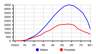
Incidence per 100.000 [ow9][d47][cym][p1e][she]
Symptoms & findings
Symptoms
Diarrhea, Fever, Hematuria, Kehr's sign, Nausea, Vomiting
Clinical findings
Elevated CRP, Hydronephrosis, Hydroureter, Leukocytosis, Perinephric stranding, Ureteral obstruction, Urinary retention
Anamneses
None listed.
Localized findings
Approach
Blood test: CBC, glucose, calcium, electrolytes (sodium, potassium, chloride, and bicarbonate), proteins (albumin and total protein), liver enzymes (ALP, ALT, and AST), bilirubin, and kidney function markers (BUN and creatinine), lactic acid, lipase, amylase, blood cultures (if febrile)
Urine stix: hematuria, leukocyte, nitrites, WBC, urine HCG
Abdominal x-ray
Ultrasound abdomen
CT abdomen
CT pelvis
Treatment
Prophylaxis: [cyu][3as]
Oral hydration that produces approximately 2.5 L of urine per day
Consumption of citrate and calcium (binds oxalate in the GI tract)
Non-operative management: 86% will pass spontaneously [cyu]
≤ 2 mm: 8 days for mean passage and passage rate of 87%
3 mm stones, 12 days for mean passage and passage rate of 76%
4-6 mm, 22 days for mean passage and passage rate of 60%
7 mm stone with a passage rate of 48%
8-9 mm stone with a passage rate of 25%
Expectant management
Analgesic: NSAIDs, Opioids reserved for refractory pain
Nausea and vomiting: Anti-emetic medications such as ondansetron, metoclopramide, promethazine
Medical expulsive therapy facilitate passage of larger (5-10 mm) stones: alpha-blockers such as doxazosin and tamsulosin
Surgical intervention to removal the stone: obstruction, oligouria, acute renal failure, infection, solitary kidney
JJ-stent for rapid renal decompression
Extracorporeal shockwave lithotripsy (ESWL): proximal stones
Flexible ureteroscopy (URS): lower pole stones between 1.5 and 2 cm
Percutaneous nephrolithotomy (PCNL): if failed ESWL/URS or >20mm proximal stones
Differential diagnoses
Abdominal abscess, Abortion, Acute mesenteric ischemia, Acute pancreatitis, Aortic dissection, Appendicitis, Cholecystitis, Constipation, Diverticulitis, Dysmenorrhea, Gastrointestinal obstruction, Glomerulonephritis, Ovarian torsion, Pelvic inflammatory disease, Pyelonephritis, Pyonephrosis, Ruptured aortic aneurysm, Ruptured ectopic pregnancy, Urinary tract infection, Xanthogranulomatous pyelonephritis
References
[1] Thakore P, Liang TH. Urolithiasis. Updated 2023 Jun 5: https://www.ncbi.nlm.nih.gov/books/NBK559101/
[2] Leslie SW, Sajjad H, Murphy PB. Renal Calculi, Nephrolithiasis. Updated 2024 Apr 20: https://www.ncbi.nlm.nih.gov/books/NBK442014/
[3] Zhang L, Zhang X, Pu Y, Zhang Y, Fan J. Global, Regional, and National Burden of Urolithiasis from 1990 to 2019: A Systematic Analysis for the Global Burden of Disease Study 2019. Clin Epidemiol. 2022 Aug 15;14:971-983. Supplementary Figure 3A
[4] Qian X, Wan J, Xu J, Liu C, Zhong M, Zhang J, Zhang Y, Wang S. Epidemiological Trends of Urolithiasis at the Global, Regional, and National Levels: A Population-Based Study. Int J Clin Pract. 2022 Mar 30;2022:6807203.
[5] GBD 2021 Urolithiasis Collaborators. The global, regional, and national burden of urolithiasis in 204 countries and territories, 2000-2021: a systematic analysis for the Global Burden of Disease Study 2021. EClinicalMedicine. 2024 Nov 21;78:102924.
[6] Li S, Huang X, Liu J, Yue S, Hou X, Hu L, Wu J. Trends in the Incidence and DALYs of Urolithiasis From 1990 to 2019: Results From the Global Burden of Disease Study 2019. Front Public Health. 2022 Mar 4;10:825541.
[7] Huang H, Li M, Fan H, Bai R. Temporal Trend of Urolithiasis Incidence in China: An Age–Period–Cohort Analysis. Int J Gen Med. 2021;14:2533-2539
[8] http://emedicine.medscape.com/article/437096 (2014-01-02); [Medscape]