Diagnosis
Pulmonary embolism
Sudden obstruction of a pulmonary artery caused by a blood clot or a substance that has travelled from elsewhere in the body through the bloodstream (embolism).
Etiology
Cause [fqu]
A complication of thrombosis in the deep venous system:
DVT from the lower extremities
Rarely: pelvic, renal, upper extremity veins or atrium
Other rare causes: Air, fat, or tumor cells embolism
Pathophysiology [fqu]
Formation of thrombi (Virchow triad):
Endothelial injury
Stasis/turbulence of blood flow
Hypercoagulability
Accretion of platelets and fibrin cause formation of thrombus
Total occlusion of the vein (Deep Venous Thrombosis)
Break off and embolize
Large saddle embolus can obstruct the main pulmonary artery and may cause:
Respiratory failure due to impaired gas exchange:
Alveolar dead space
Ventilation-perfusion mismatch
Pulmonary infarction (intra-alveolar hemorrhage)
Hemodynamic compromise
Increased pulmonary vascular resistance
Increased right ventricular afterload
Reduced cardiac output
Classification of pulmonary embolism:
Hemodynamically stable PE: mildly symptomatic, or asymptomatic
Hemodynamically unstable PE: risk of right ventricular failure and shock
Systolic blood pressure <90 mm Hg
Drop in systolic blood pressure of ≥40 mm Hg from baseline
Hypotension that requires vasopressors or inotropes
Risk factors [fqu][hpm]
Hereditary risk factors: Antithrombin III deficiency, Protein C deficiency, Protein S deficiency, Factor V Leiden, Plasminogen abnormality, Plasminogen activator abnormality, Fibrinogen abnormality, Resistance to activated protein C
Acquired risk factors: Old age, cancer, immobilization (paresis, heart failure, respiratory failure, trauma, fracture), smoking, recent surgery, pregnancy, hormone replacement therapy, oral contraceptive therapy, fibrillation/flutter, central venous lines, chemotherapy, infection, obesity
High-risk Pulmonary Embolism: Shock, hypotension, CT/echocardiography (RV dilatation, hypokinesis or pressure overload), elevated BNP/proBNP, elevated cardiac troponin, right heart catheterization (Elevated right heart pressure)
Complications [fqu]
Recurrent thromboembolism
Chronic thromboembolic pulmonary hypertension
Right heart failure
Cardiogenic shock
Epidemiology
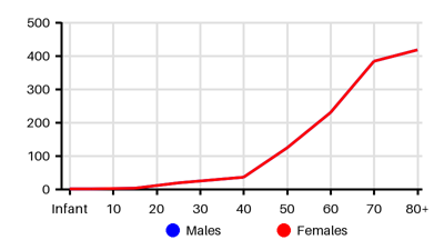
Incidence per 100.000 [dfv][too][pyr][fqu][5rs][hpm][pvf][so1][udu][bac][4id][nyf][cad][q9g][bzf][sis][8em]
Symptoms & findings
Symptoms
Cough, Crepitations, Cyanosis, Decreased consciousness, Delirium, Diaphoresis, Dyspnea, Edema, Fever, Heart murmur, Hemoptysis, Hypotension, Jugular vein distention, Oliguria, Seizure, Syncope, Tachycardia, Tachypnea, Third heart sound, Thrombophlebitis, Wheezing
Clinical findings
Atrial fibrillation, Elevated BNP, Elevated D-dimer, Elevated Troponin, Friction rub, Hypoxemia, Leukocytosis, Pleural effusion, Pleuritis, Pulsus paradoxus, Shock
Anamneses
Estrogen treatment, Immobilization, Malignancy, Pregnancy, Smoking, Surgery, Trauma
Localized findings
Approach
Blood test: [hpm][fqu]
Troponin, BNP, D-dimer (high negative predictive value in patients with a low or moderate clinical probability)
Blood gas
ECG
Echocardiography: right ventricule overload
Chest x-ray
CT angiography
V/Q scanning
Venography
Duplex ultrasonography: Compression of the deep veins
Flowchart for diagnosis [hpm]
Suspected non-high-risk Pulmonary Embolism -> Clinical score (Wells/Geneva)
Low/intermediate clinical probability -> D-dimer:
Negative D-dimer -> No treatment
Positive D-dimer -> CT
High clinical probability -> CT:
No PE -> No treatment (investigate further)
Suspected high-risk Pulmonary Embolism: Right Ventricule failure -> shock
Stable patient -> CT
Unstable patient -> Echocardiography
Geneva score [d7k]
Predisposing factors
+1 Age > 65years
+3 Previous DVT or PE
+2 Surgery or fracture within 1 month
+2 Active malignancy
Symptoms
+3 Unilateral lower limb pain
+2 Hemoptysis
Clinical signs
+3 heart rate 75-94 beats/min
+5 heart rate ≥95 beats/min
+4 Pain on palpation and unilateral edema on lower limb
Clinical probability:
0-3 Low (~10%)
4-10 Intermediate (~30%)
≥11 High (~65%)
Wells score [s6c]
Predisposing factors
+1.5 Previous DVT or PE
+1.5 Recent surgery or immobilization
+1 Cancer
Symptoms
+1 Hemoptysis
Clinical signs
+1.5 heart rate >100 beats/min
+3 Clinical signs of DVT
+3 Alternative diagnosis less likely than PE
Clinical probability:
0-1 Low (10%)
2-6 Intermediate (~30%)
≥7 High (~65%)
Treatment
Hemodynamically stable PE [hpm]
Enoxaparin 1.0 mg/kg Every 12 h or 1.5 mg/kga Once daily
Warfarin: INR of 2.5 (range 2.0–3.0) duration of 3–6 months
Hemodynamically unstable PE [hpm]
Oxygen:
Increase supply: nasal/mechanical ventilation without PEEP to maximize preload
Decrease consumption (reduce fever and agitation)
Modest fluid resucitation
Inotropic drugs (Isoproterenol, Norepinephrine, Dobutamine, Dopamine)
Vasodilators administered by inhalation (nitric oxide) decrease pulmonary vascular resistance
Anticoagulation:
Enoxaparin 1.0 mg/kg Every 12 h or 1.5 mg/kga Once daily
Thrombolysis: contraindicated of stroke/brain hemorrhage, brain cancer, major trauma/surgery. Relative contraindication: pregnancy, liver failure, endocarditis, ulcer, traumatic resuscitation
Recombinant tissue plasminogen activator (rtPA) 100 mg over 2 h or 0.6 mg/kg over 15 min (maximum dose 50 mg)
Streptokinase 250 000 IU as a loading dose over 30 min, followed by 100 000 IU/h over 12– 24 h. Accelerated regimen: 1.5 million IU over 2 h
Urokinase 4400 IU/kg as a loading dose over 10 min, followed by 4400 IU/kg/h over 12– 24 h. Accelerated regimen: 3 million IU over 2 h
Embolectomy (endovascular/surgical):
Alternative to thrombolysis when there are absolute contraindications
Adjunctive therapy when thrombolysis has failed to improve hemodynamics
Vena cava filter: Retrievable filters may be considered in selected patients with recurrent venoius thromboembolism (DVT or PE) despite anticoagulation or in patients with absolute contraindication to anticoagulants [fqu]
Differential diagnoses
Acute respiratory distress syndrome, Allergic alveolitis, Angina pectoris, Anxiety, Aortic stenosis, Arrhythmia, Atrial fibrillation, Cardiac tamponade, Cardiomyopathy, COPD, Cor pulmonale, Emphysema, Fat embolism syndrome, Heart failure, Hyperventilation, Mediastinitis, Mitral stenosis, Myocardial infarction, Pericarditis, Pleuritis, Pneumonia, Pneumonitis, Pneumothorax, Pulmonary edema, Pulmonary hypertension, Shock, Sickle cell disease, Superior vena cava syndrome, Syncope, Vasovagal syncope
References
[1] Vyas V, Sankari A, Goyal A. Acute Pulmonary Embolism. Updated 2024 Dec 11: https://www.ncbi.nlm.nih.gov/books/NBK560551/
[2] Torbicki A, Perrier A, Konstantinides S, Agnelli G, Galiè N, Pruszczyk P, Bengel F, Brady AJ, Ferreira D, Janssens U, Klepetko W, Mayer E, Remy-Jardin M, Bassand JP; ESC Committee for Practice Guidelines (CPG). Guidelines on the diagnosis and management of acute pulmonary embolism: the Task Force for the Diagnosis and Management of Acute Pulmonary Embolism of the European Society of Cardiology (ESC). Eur Heart J. 2008 Sep;29(18):2276-315.
[3] Venous thromboembolism: incidence and risk factors. Nat Rev Cardiol 4, 468 (2007).
[4] Nordstrom M, Lindblad B. Autopsy-verified venous thromboembolism within a defined urban population - the city of Malmo, Sweden. APMIS 1998;106: 378 –384.
[5] Oger E. Incidence of venous thromboembolism: a community-based study in Western France. EPI-GETBP Study Group. Groupe d’Etude de la Thrombose de Bretagne Occidentale. Thromb Haemost 2000;83:657 –660.
[6] Meignan M, Rosso J, Gauthier H, Brunengo F, Claudel S, Sagnard L, d'Azemar P, Simonneau G, Charbonnier B. Systematic lung scans reveal a high frequency of silent pulmonary embolism in patients with proximal deep venous thrombosis. Arch Intern Med. 2000 Jan 24;160(2):159-64.
[7] Silverstein MD, Heit JA, Mohr DN, Petterson TM, O'Fallon WM, Melton LJ 3rd. Trends in the incidence of deep vein thrombosis and pulmonary embolism: a 25-year population-based study. Arch Intern Med. 1998 Mar 23;158(6):585-93.
[8] Anderson FA Jr, Wheeler HB, Goldberg RJ, Hosmer DW, Patwardhan NA, Jovanovic B, Forcier A, Dalen JE. A population-based perspective of the hospital incidence and case-fatality rates of deep vein thrombosis and pulmonary embolism. The Worcester DVT Study. Arch Intern Med. 1991 May;151(5):933-8.
[9] Stein PD, Huang Hl, Afzal A, Noor HA. Incidence of acute pulmonary embolism in a general hospital: relation to age, sex, and race. Chest. 1999 Oct;116(4):909-13.
[10] Proctor MC, Greenfield LJ. Pulmonary embolism: diagnosis, incidence and implications. Cardiovasc Surg. 1997 Feb;5(1):77-81.
[11] Cheuk BL, Cheung GC, Cheng SW. Epidemiology of venous thromboembolism in a Chinese population. Br J Surg. 2004 Apr;91(4):424-8.
[12] Wendelboe AM, Raskob GE. Global Burden of Thrombosis: Epidemiologic Aspects. Circ Res. 2016 Apr 29;118(9):1340-7.
[13] Centers for Disease Control and Prevention (CDC). Venous thromboembolism in adult hospitalizations - United States, 2007-2009. MMWR Morb Mortal Wkly Rep. 2012 Jun 8;61(22):401-4.
[14] Jang MJ, Bang SM, Oh D. Incidence of venous thromboembolism in Korea: from the Health Insurance Review and Assessment Service database. J Thromb Haemost. 2011 Jan;9(1):85-91.
[15] Sonne-Holm E, Kjærgaard J, Bang LE, Fosbøl E, Carlsen J, Winther-Jensen M. Pulmonary embolism: Age specific temporal trends in incidence and mortality in Denmark 1999-2018. Thromb Res. 2022 Feb;210:12-19.
[16] Lehnert P, Lange T, Møller CH, Olsen PS, Carlsen J. Acute Pulmonary Embolism in a National Danish Cohort: Increasing Incidence and Decreasing Mortality. Thromb Haemost. 2018 Mar;118(3):539-546.
[17] Heit JA. Epidemiology of venous thromboembolism. Nat Rev Cardiol. 2015 Aug;12(8):464-74. doi: 10.1038/nrcardio.2015.83. Epub 2015 Jun 16.
[18] Le Gal G, Righini M, Roy PM, Sanchez O, Aujesky D, Bounameaux H et al. Prediction of pulmonary embolism in the emergency department: the revised Geneva score. Ann Intern Med 2006;144:165– 171
[19] Wells PS, Anderson DR, Rodger M, Ginsberg JS, Kearon C, Gent M et al. Derivation of a simple clinical model to categorize patients probability of pulmonary embolism: increasing the models utility with the SimpliRED D-dimer. Thromb Haemost 2000;83:416 –420.
[20] http://emedicine.medscape.com/article/300901 (2014-01-03); [Medscape]