Diagnosis
Myocardial infarction
Pathological definition: The irreversible death (necrosis) of heart muscle secondary to prolonged lack of oxygen supply (ischemia)
Clinical definition: [g0z]
Charactiristic chest pain
Elevation of cardiac enzymes (Troponin, CK-MB)
Electrocardiographic (ECG) changes: ST-segment, Q wave
Radiology (ultrasound, angiography) detecting myocardial injury and necrosis
Also known as: Heart attack
Etiology
Causes [g0z]
Type 1: Spontaneous
Plaque rupture
Ulceration
Fissuring
Erosion
Aortic dissection with involvement of ostia --> intraluminal thrombus
Type 2: Secondary to increased oxygen demand or a decreased supply
Coronary artery spasm
Vasculitis
Congenital coronary anomalies
Coronary artery embolus
Tachyarryhthmias/bradyarrhythmias
Anemia
Respiratory failure
Hypertension
Hypotension
Type 3: Sudden cardiac death without available blood sample for biomarkers
Type 4a: Related to PCI
Type 4b: Related to stent thrombosis
Type 5: Related to coronary artery bypass grafting
Pathophysiology [yzw]
Ischemia = supply of oxygen < demand of oxygen
Preload
Afterload
Wall thickness
Heart rate
Contractility
Atherosclerotic plaque --> stenosis --> impedes flow
Cross-sectional area is reduced by 75% = 50% reduction in diameter
Intermittent closure of dynamic plaques --> unstable angina
Thrombosis or plaque rupture --> occlusion >20 minutes --> myocardial infarction
Depending on the territory and the thickness affected by the infarction, the cardiac function is compromised:
Unstable angina: cardiac chest pain + normal ECG/biomarkers
NSTEMI: cardiac chest pain + elevated cardiac biomarker levels
STEMI: chest pain + ST-segment elevation + elevated cardiac biomarker levels
Negligible regeneration capacity --> scar formation
Remodeling: dilation, segmental hypertrophy of remaining viable tissue, and cardiac dysfunction
Complications
Malignant ventricular arrhythmia
Ventricular wall rupture
Ventricular septal defect
Papillary muscle rupture with severe mitral regurgitation
Ventricular aneurysm formation
Risk factors [yzw]
Hypertension
Smoking
Hypercholesterolemia
Obesity
Diabetes mellitus
Family history
Epidemiology
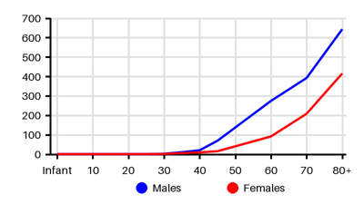
Incidence per 100.000 [pwn][63h][6ct][yqb]
Symptoms & findings
Symptoms
Anxiety, Arrhythmia, Diaphoresis, Diastolic murmur, Distant heart sounds, Dizziness, Dyspnea, Edema, Fatigue, Heart murmur, Malaise, Nausea, Oliguria, Palpitations, Syncope, Systolic murmur, Tachycardia, Weakness, Wheezing
Clinical findings
Decreased Ejection Fraction, Elevated Troponin, Inverted T wave, Mitral regurgitation, Myocardial hypokinesia, Pathologic Q wave, Pericardial effusion, Pleural effusion, Pulmonary edema, Pulsus paradoxus, Shock, ST depression, ST elevation
Anamneses
None listed.
Localized findings
Approach
Blood test: Troponin, CK-MB, lipid profile, complete blood cell count
ECG
Stress test
Echocardiography
Angiography
Chest x-ray
Treatment
Prehospital care: [g0z]
Intravenous access
Pulse oximetry --> Supplemental oxygen if SaO2 < 90%
Aspirin 300mg
Nitroglycerin 0.4mg sublingually/spray
Pain: Morphine 2-4 mg IV repeated every 5-10 minutes until relieved
Telemetry and prehospital ECG
STEMI:
<90 minutes: emergent coronary angiography, primary PCI, drug-eluting stent
>120 minutes from a PCI-capable facility --> fibrinolysis
Coronary artery bypass grafting: cardiogenic shock, failed PCI, high-risk anatomy
NSTEMI:
Anti-ischemic therapy
May be candidates for PCI at admission
Treatment on long term:
Anticoagulants
Antiplatelet agents
Beta blockers
Statins
Angiotensin-converting enzyme (in some patients)
Lifestyle modifications
Differential diagnoses
Abdominal compartment syndrome, Angina pectoris, Anxiety, Aortic dissection, Aortic regurgitation, Aortic stenosis, Arrhythmia, Asthma, Cholangitis, Cholecystitis, Cholelithiasis, COPD, Depression, Endocarditis, Esophageal spasm, Esophagitis, Gastritis, Gastroesophageal reflux disease, Herpes zoster, Mitral regurgitation, Myocarditis, Pericarditis, Pleuritis, Pneumonia, Pneumothorax, Pulmonary embolism, Ruptured aortic aneurysm, Shock
References
[1] http://emedicine.medscape.com/article/155919 (2014-01-02); [Medscape]
[2] Ojha N, Dhamoon AS. Myocardial Infarction. [Updated 2023 Aug 8]. In: StatPearls [Internet]. Treasure Island (FL): StatPearls Publishing; 2025 Jan-. Available from: https://www.ncbi.nlm.nih.gov/books/NBK537076/
[3] Christensen DM, Strange JE, Phelps M, Schjerning AM, Sehested TSG, Gerds T, Gislason G. Age- and sex-specific trends in the incidence of myocardial infarction in Denmark, 2005 to 2021. Atherosclerosis. 2022 Apr;346:63-67
[4] Reynolds K, Go AS, Leong TK, Boudreau DM, Cassidy-Bushrow AE, Fortmann SP, Goldberg RJ, Gurwitz JH, Magid DJ, Margolis KL, McNeal CJ, Newton KM, Novotny R, Quesenberry CP Jr, Rosamond WD, Smith DH, VanWormer JJ, Vupputuri S, Waring SC, Williams MS, Sidney S. Trends in Incidence of Hospitalized Acute Myocardial Infarction in the Cardiovascular Research Network (CVRN). Am J Med. 2017 Mar;130(3):317-327
[5] Yamamoto T, Yoshida N, Takayama M; Tokyo CCU Network. Temporal Trends in Acute Myocardial Infarction Incidence and Mortality Between 2006 and 2016 in Tokyo - Report From the Tokyo CCU Network. Circ J. 2019 May 24;83(6):1405-1409
[6] Kuhn J, Olié V, Grave C, Le Strat Y, Bonaldi C, Joly P. Estimating the Future Burden of Myocardial Infarction in France Until 2035: An Illness-Death Model-Based Approach. Clin Epidemiol. 2022 Mar 5;14:255-264