Diagnosis
Pneumothorax
Presence of air in the pleural cavity (space between the visceral and parietal pleura).
Etiology
Cause [f9e][hne]
Primary spontaneous pneumothorax: No underlying lung disease or trauma
Subclinical pleural blebs/bullae that may rupture and cause pneumothorax
Risk factors: pregnancy, tall, thin, male, smoker, age 18-40 years, Marfan syndrome
Seconday spontaneous pneumothorax: Underlying pulmonary pathology
Risk factors: COPD, asthma, HIV, pneumonia, tuberculosis, sarcoidosis, cystic fibrosis, lung cancer, pulmonary fibrosis, SARS, endometriosis (catamenial pneumothorax), Langerhans cell histiocytosis, lymphangioleiomyomatosis, connective tissue diseases (rheumatoid arthritis, scleroderma, Marfan syndrome, Ehlers-Danlos syndrome, ankylosing spondylitis, polymyositis/dermatomyositis)
Iatrogenic pneumothorax: thoracentesis, central venous catheter, tracheostomy, transbronchial biopsy, CT–guided needle lung biopsy, positive pressure ventilation (barotrauma), intercostal nerve block
Traumatic pneumothorax: blunt trauma (rib fracture), penetrating trauma, diving or flying
Pathophysiology [f9e]
Ventilation:
The pressure of the pleural space is negative compared to atmospheric pressure
The chest wall expands outwards --> the lung also expands outwards --> Air fills inside the lungs
Pneumothorax: Air in the pleural cavity can result in a collapse of the lung and impair oxygenation and/or ventilation
Tension pneumothorax: A life-threatening condition where air under positive pressure is trapped in the pleural cavity and the injured tissue forms a 1-way valve, allowing air inflow with inhalation into the pleural space and prohibiting air outflow. As the pressure increases --> ipsilateral lung collapses --> mediastinal shift --> contralateral lung collapse --> impaired venous return due to kinking of the inferior vena cava --> acute heart failure
Most common causes: blunt/penetrating trauma, positive pressure ventilation, pneumoperitoneum, bronchoscopy, tracheostomy
Complications [f9e]
Respiratory failure
Cardiac arrest
Pyopneumothorax
Empyema
Rexpansion pulmonary edema
Pneumopericardium
Pneumoperitoneum
Pneumohemothorax
Bronchopulmonary fistula
Hemorrhage during tube thoracostomy
Skin infection at the site of tube thoracostomy
Epidemiology
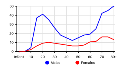
Incidence per 100.000 [wdr][cy3][dpy][kis][f9e][g4s][dsr]
Symptoms & findings
Symptoms
Anxiety, Cough, Crepitations, Cyanosis, Decreased consciousness, Decreased respiratory sounds, Diaphoresis, Dyspnea, Fatigue, Hamman's sign, Hypotension, Jugular vein distention, Malaise, Respiratory distress, Respiratory failure, Subcutaneous emphysema, Tachycardia, Tachypnea, Tracheal deviation
Clinical findings
Acidosis, Elevated Jugular Venous Pressure, Hypoxemia, Mediastinal shift, Pneumomediastinum, Pulsus paradoxus
Anamneses
None listed.
Localized findings
Approach
Blood gas
Chest x-ray
CT thorax
Contrast esophagography
Chest ultrasonography
Fluorescein-enhanced autofluorescence thoracoscopy
Treatment
Signs of instability --> needle decompression: [f9e]
14- to 16-gauge and 4.5 cm in length angiocatheter
Flush above the rib in the 2nd intercostal space in the midclavicular line
Proceed with tube thoracostomy
Stable patient:
If asymptomatic and depth/size 1-2cm --> Watchful waiting or needle aspiration
If symptomatic or the depth/size >2cm --> Tube thoracostomy
Flush above the rib in the fifth intercostal space anterior to the midaxillary line
Indication for pleurodesis or bullectomy: VATS vs. thoracotomy vs laser treatment
Continuous air leak for >7 days
Bilateral pneumothoraces
High-risk profession patients (divers, pilots)
Recurrent ipsilateral pneumothorax
Contralateral pneumothorax
Patients who have AIDS
Open chest wounds:
Damage control: Three-sided occlusive dressing
Definitive surgery: Tube thoracostomy --> chest wall defect repair
Differential diagnoses
Acute respiratory distress syndrome, Aortic dissection, Asthma, Cardiac tamponade, COPD, Costochondritis, Esophageal perforation, Esophageal spasm, Fracture, Heart failure, Hemothorax, Lung abscess, Mediastinitis, Myocardial infarction, Myocarditis, Pericarditis, Pleural effusion, Pleuritis, Pneumomediastinum, Pneumonia, Pulmonary edema, Pulmonary embolism, Pulmonary empyema, Rib fracture, Tuberculosis
References
[1] McKnight CL, Burns B. Pneumothorax. Updated 2023 Feb 15: https://www.ncbi.nlm.nih.gov/books/NBK441885/
[2] Noppen M. Spontaneous pneumothorax: epidemiology, pathophysiology and cause. Eur Respir Rev. 2010 Sep;19(117):217-9.
[3] Hallifax RJ, Goldacre R, Landray MJ, Rahman NM, Goldacre MJ. Trends in the Incidence and Recurrence of Inpatient-Treated Spontaneous Pneumothorax, 1968-2016. JAMA. 2018 Oct 9;320(14):1471-1480.
[4] Bobbio A, Dechartres A, Bouam S, Damotte D, Rabbat A, Régnard JF, Roche N, Alifano M. Epidemiology of spontaneous pneumothorax: gender-related differences. Thorax. 2015 Jul;70(7):653-8.
[5] Zhong X, Goldacre R, Morris EJA, Hallifax RJ. Trends in incidence of pneumothorax in England before, during and after the COVID-19 pandemic (2017-2023): a population-based observational study. Lancet Reg Health Eur. 2024 Jul 1;44:100994.
[6] Huang YH, Chang PY, Wong KS, Chang CJ, Lai JY, Chen JC. An Age-Stratified Longitudinal Study of Primary Spontaneous Pneumothorax. J Adolesc Health. 2017 Oct;61(4):527-532.
[7] Melton LJ 3rd, Hepper NG, Offord KP. Incidence of spontaneous pneumothorax in Olmsted County, Minnesota: 1950 to 1974. Am Rev Respir Dis. 1979 Dec;120(6):1379-82.
[8] Olesen WH, Titlestad IL, Andersen PE, Lindahl-Jacobsen R, Licht PB. Incidence of primary spontaneous pneumothorax: a validated, register-based nationwide study. ERJ Open Res. 2019 Jun 10;5(2):00022-2019.
[9] http://emedicine.medscape.com/article/424547 (2014-01-04); [Medscape]