Diagnosis
Pleural effusion
Abnormal collection of fluid in the pleural space (between parietal and visceral pleura) resulting from excess fluid production or decreased absorption.
Etiology
Cause [twi]
75% of the cases: Heart failure, cancer, pneumonia, and tuberculosis [r3t]
Transudate: low protein due to imbalance in hydrostatic and oncotic forces
Heart failure, cirrhosis, decreased albumin, atelectasis (due to malignancy or pulmonary embolism), nephrotic syndrome, peritoneal dialysis, constrictive pericarditis, urinothorax, cerebrospinal fluid (ventriculopleural shunting or thoracic spine surgery), extravascular migration of central venous catheter
Exudate: high protein in areas of inflammation due to increased capillary permeability
Bacterial pneumonia, lung or breast cancer, lymphoma, leukemia; less commonly, ovarian carcinoma, stomach cancer, sarcomas, melanoma), pulmonary embolism, rheumatoid arthritis, systemic lupus erythematosus, tuberculosis, pancreatitis, trauma, esophageal perforation, radiation pleuritis, sarcoidosis, fungal infection, pancreatic pseudocyst, abdominal abscess, pericardial disease, Meigs syndrome, ovarian hyperstimulation syndrome, drug side effects (methotrexate, amiodarone, phenytoin, and dasatinib), yellow nail syndrome, renal failure, chylothorax, pseudochylothorax, fistula
Pathophysiology [twi]
Normal pleural fluid serves as a lubricant to facilitate movement of the pleural surfaces against each other in the course of respirations
This small volume of fluid is maintained through the balance of hydrostatic and oncotic pressure and lymphatic drainage
Exudate:
Altered pleural permeability: pneumonia, malignancy, pulmonary embolism
Increased capillary permeability: trauma, malignancy, inflammation, pulmonary embolism, drug hypersensitivity, renal failure, pancreatitis
Transudate:
Decreased intravascular oncotic pressure: decreased albumin, cirrhosis
Increased capillary hydrostatic pressure: heart failure, vena cava syndrome
Decreased pressure in the pleural space (preventing full lung expansion): atelectasis, mesothelioma
Decreased lymphatic drainage: malignancy, trauma
Transudation from ascites or pulmonary edema
The net result may be compression of the lungs --> restrictive ventilatory defect:
Dyspnea
Chest pain
Cough
Complications [twi]
Empyema
Sepsis
Respiratory compromise
Pleural thickening --> restrictive lung disease
Epidemiology
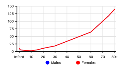
Incidence per 100.000 [ksd][pzp][yms][nuy][4kb][i4p][sab]
Symptoms & findings
Symptoms
Anorexia, Cough, Decreased respiratory sounds, Dyspnea, Edema, Fever, Hemoptysis, Orthopnea, Tracheal deviation
Clinical findings
Anamneses
None listed.
Localized findings
Approach
Blood test: WBC, CRP, NT-proBNP
Blood gas
Chest x-ray
CT thorax
Ultrasound
Thoracentesis: pH, glucose, protein, LD, amylase, cholesterol, ANA, RF, differential cell counts, Gram stain, culture, cytology and biopsy
Light’s criteria to distinguish transudates and exudates [twi]
Exudative meets ≥1 of the following criteria; a transudate meets none of the criteria:
Pleural fluid protein/serum protein >0.5
Pleural fluid LDH/serum lactate dehydrogenase (LDH) >0.6
Pleural fluid LDH >2/3rds of the upper normal limit of serum LDH
Heffner's criteria for exudative effusion [twi]
Pleural fluid protein level exceeding 2.9 g/dL
Pleural fluid cholesterol level greater than 45 mg/dL
Pleural LDH level greater than two-thirds of the upper limit of normal serum LDH
Pleural effusion analysis
Pleural fluid:
pH 7.60-7.64
Protein 1-2 g/dl
WBC < 1000
Lactate Dehydrogenase 50% of plasma
Pleural LD > 1000 IU/L:
Empyema
Malignant effusion
Rheumatoid effusion
Pleural paragonimiasis.
Low pleural glucose/pH:
Malignant effusion
Tuberculous pleuritis
Esophageal rupture
Lupus pleuritis
Pleural lymphocytosis:
TB
Lymphoma
Sarcoidosis
Chronic rheumatoid pleurisy
Yellow nail syndrome
Chylothorax
Malignancy
Treatment
Treating the underlying cause [hdn]
Goals:
Sterilization of pleural fluid
Reexpansion of the lung
Restoration of normal lung function
Antibiotics
Thoracocentesis: compromising respiratory function
Pleural drainage
Chest tube drainage with or without instillation of fibrinolytic agents
Surgery:
Video-assisted thoracoscopic surgery
Open thoracotomy with decortication
Differential diagnoses
Abdominal abscess, Acute pancreatitis, Acute renal failure, Acute respiratory distress syndrome, Amyloidosis, Atelectasis, Breast cancer, Chronic renal failure, Chylothorax, Crohn disease, Esophageal perforation, Esophageal rupture, Heart failure, Hemothorax, Hypothyroidism, Leukemia, Lung cancer, Lymphoma, Meigs syndrome, Mesothelioma, Nephrotic syndrome, Pericarditis, Peritonitis, Pneumonia, Pulmonary edema, Pulmonary embolism, Renal failure, Rheumatoid arthritis, Sepsis, Superior vena cava syndrome, Systemic lupus erythematosus, Tuberculosis, Ulcerative colitis, Yellow nail syndrome
References
[1] Krishna R, Antoine MH, Alahmadi MH, et al. Pleural Effusion. [Updated 2024 Aug 31]. In: StatPearls [Internet]. Treasure Island (FL): StatPearls Publishing; 2025 Jan-. Available from: https://www.ncbi.nlm.nih.gov/books/NBK448189/
[2] Botana Rial M, Pérez Pallarés J, Cases Viedma E, López González FJ, Porcel JM, Rodríguez M, Romero Romero B, Valdés Cuadrado L, Villena Garrido V, Cordovilla Pérez R. Diagnosis and Treatment of Pleural Effusion. Recommendations of the Spanish Society of Pulmonology and Thoracic Surgery. Update 2022. Arch Bronconeumol. 2023 Jan;59(1):27-35. English, Spanish.
[3] Mummadi SR, Stoller JK, Lopez R, Kailasam K, Gillespie CT, Hahn PY. Epidemiology of Adult Pleural Disease in the United States. Chest. 2021 Oct;160(4):1534-1551.
[4] Sorg AL, Obermeier V, Liese JG, von Kries R. Incidence trends of parapneumonic pleural effusions/empyema in children 2009 to 2018 from health insurance data: Only temporal reduction after the introduction of PCV13. Vaccine. 2021 Jun 11;39(26):3516-3519.
[5] Wiese AD, Griffin MR, Zhu Y, Mitchel EF Jr, Grijalva CG. Changes in empyema among U.S. children in the pneumococcal conjugate vaccine era. Vaccine. 2016 Dec 7;34(50):6243-6249.
[6] Byington CL, Spencer LY, Johnson TA, Pavia AT, Allen D, Mason EO, Kaplan S, Carroll KC, Daly JA, Christenson JC, Samore MH. An epidemiological investigation of a sustained high rate of pediatric parapneumonic empyema: risk factors and microbiological associations. Clin Infect Dis. 2002 Feb 15;34(4):434-40.
[7] Byington CL, Korgenski K, Daly J, Ampofo K, Pavia A, Mason EO. Impact of the pneumococcal conjugate vaccine on pneumococcal parapneumonic empyema. Pediatr Infect Dis J. 2006 Mar;25(3):250-4.
[8] Deceuninck G, Quach C, Panagopoulos M, Thibeault R, Côté-Boileau T, Tapiéro B, Coïc L, De Wals P, Ovetchkine P. Pediatric Pleural Empyema in the Province of Quebec: Analysis of a 10-Fold Increase Between 1990 and 2007. J Pediatric Infect Dis Soc. 2014 Jun;3(2):119-26.
[9] Shin EJ, Kim Y, Jeong JY, Jung YM, Lee MH, Chung EH. The changes of prevalence and etiology of pediatric pneumonia from National Emergency Department Information System in Korea, between 2007 and 2014. Korean J Pediatr. 2018 Sep;61(9):291-300.
[10] https://emedicine.medscape.com/article/1003121
[11] http://emedicine.medscape.com/article/299959 (2014-01-06); [Medscape]
[12] Mahon C, Walker W, Drage A, Best E. Incidence, aetiology and outcome of pleural empyema and parapneumonic effusion from 1998 to 2012 in a population of New Zealand children. J Paediatr Child Health. 2016 Jun;52(6):662-8.