Diagnosis
Pulmonary edema
An abnormal accumulation of extravascular fluid in the lung parenchyma causing diminished gas exchange at the alveolar level and respiratory failure.
Etiology
Cause [dqo]
Cardiogenic Pulmonary Edema: Acute cardiac event -> elevated pulmonary venous pressure -> increased hydrostatic pressure -> transudation of fluid into the pulmonary interstitium/alveoli -> decreased alveolar gas exchange -> respiratory failure
Excessive intravascular volume administration
Congestive left-sided heart failure
Valvular heart disease (mitral stenosis or aortic stenosis)
Atrial fibrillation
Myocardial infarction
Cardiomyopathy
Myxoma
Hypertensive crisis
Pulmonary embolism with associated right heart strain
Right to left shunts
Non-Cardiogenic Pulmonary Edema: Lung injury -> increased pulmonary capillary permeability
Acute respiratory distress syndrome (ARDS)
Pneumonia or sepsis/anaphylactic shock
Toxin inhalation (smoke, chlorine gas)
High-altitude pulmonary edema (HAPE)
Neurogenic pulmonary edema (after head trauma or seizures)
Drug overdose (opioids, aspirin toxicity)
Acute pancreatitis
Blood transfusion-related lung injury (TRALI)
Pulmonary contusion
Radiation
Drowning
Fat emboli
Other:
Lymphatic obstruction due to lymphoma, fibrosing lymphangitis or lung transplantation: The lymphatics play an important role in maintaining an adequate fluid balance in the lungs by removing solutes, colloid, and liquid from the interstitial space.
Neurogenic pulmonary edema (neurological events associated with elevated intracranial pressure) and high-altitude pulmonary edema.
Thyrotoxicosis
Myocarditis
Myocardial toxins (alcohol, cocaine, chemotherapeutic agents)
Pathophysiology [dqo][rh5]
The alveolar gas and capillary blood is separated by the alveolar-capillary membrane (capillary endothelium, interstitial space and alveolar epithelium)
Pulmonary edema occurs when there is increased transudation of fluid into the pulmonary interstitium and alveoli -> alveolar flooding -> severe respiratory failure
Disruption of the balance of fluid filtration:
Increase in intravascular hydrostatic pressure (pulmonary vein pressure)
Increase in interstitial hydrostatic pressure
Endothelial injury and disruption of epithelial barriers
Decrease in oncotic pressure (decreased albumin: hepatic, renal, malnutrition)
Lymphatic insufficiency
Increased negative interstitial pressure
Complications [dqo]
Respiratory failure
ARDS
Cardiac complications
Multiorgan failure
Epidemiology
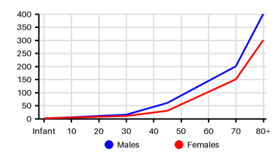
Incidence per 100.000 [rh5][efo]
Symptoms & findings
Symptoms
Anxiety, Confusion, Cough, Crepitations, Diaphoresis, Diastolic murmur, Dizziness, Dyspnea, Fatigue, Heart murmur, Hoarseness, Jugular vein distention, Orthopnea, Pallor, Sputum, Systolic murmur, Tachycardia, Tachypnea, Third heart sound, Weight gain
Clinical findings
Anemia, Elevated BNP, Elevated Jugular Venous Pressure, Hypercapnia, Hypoxemia, Leukocytosis
Anamneses
None listed.
Localized findings
Approach
Blood test: Hb, WBC, CRP, electrolytes, brain natriuretic peptide, troponin, albumin
Blood gas
Blood culture
Edema fluid-to-plasma protein ratio [sqy]
ECG
Chest x-ray
Echocardiography
Lung ultrasound
CT thorax
Pulmonary Arterial Catheter
Treatment
Initial symptomatic treatment: [dqo]
Airways: Suction of mucus
Breathing: Oxygen, nasal cannula, face mask, noninvasive/invasive ventilatory support, intubation
Circulation: Inotropes if hypotension (Dobutamine, Dopamine, Milrinone)
Diuretics: Furosemide 20 to 40 mg IV, bumetanide 1 mg IV
Vasodilators for Pre-Load Reduction if systolic blood pressure > 110 mm Hg:
IV nitroglycerin lowers preload and pulmonary congestion
Sodium nitroprusside
Isosorbide dinitrate
Nifedipine
Nesiritide (with care)
Morphine (analgesic/anxiolytic): with care due to respiratory depression
Ultrafiltration: Sodium and water removal
Monitor: Blood pressure, respiratory rate, pulse, urine output, pedal edema, weight, intake/output
Treatment of the underlying condition
Differential diagnoses
Acute liver failure, Acute respiratory distress syndrome, Anaphylaxis, Asthma, COPD, Emphysema, Goodpasture syndrome, Myocardial infarction, Pneumonia, Pneumothorax, Pulmonary embolism, Renal failure, Respiratory failure, Shock, Stroke
References
[1] Malek R, Soufi S. Pulmonary Edema. Updated 2023 Apr 7: https://www.ncbi.nlm.nih.gov/books/NBK557611/
[2] Iqbal MA, Gupta M. Cardiogenic Pulmonary Edema. Updated 2023 Apr 7: https://www.ncbi.nlm.nih.gov/books/NBK544260/
[3] Edoute Y, Roguin A, Behar D, Reisner SA. Prospective evaluation of pulmonary edema. Crit Care Med. 2000 Feb;28(2):330-5.
[4] Ware LB, Fremont RD, Bastarache JA, Calfee CS, Matthay MA. Determining the aetiology of pulmonary oedema by the oedema fluid-to-plasma protein ratio. Eur Respir J. 2010 Feb;35(2):331-7.
[5] http://emedicine.medscape.com/article/300716 (2014-01-02); [Medscape]
[6] http://emedicine.medscape.com/article/157452 (2014-01-02); [Medscape]
[7] Platz E, Jhund PS, Campbell RT, McMurray JJ. Assessment and prevalence of pulmonary oedema in contemporary acute heart failure trials: a systematic review. Eur J Heart Fail. 2015 Sep;17(9):906-16.