Diagnosis
Cardiac tamponade
Accumulation of fluid in the pericardial space, resulting in reduced ventricular filling and subsequent hemodynamic compromise.
Also known as: Pericardial tamponade
Etiology
Cause [74u]
Cancer --> pleural effusion (>50% of cases)
Lung >70%
Breast
Renal
Lymphoma
Leukemia
Uremia - 10-15%
Pericarditis - 5-15%
Infectious diseases - 5-10%
Staphylococcus aureus
Myobacterium tuberculosis
Staphylococcus pneumonia
HIV
Coxsackievirus group B
Invluenza
Echoviruses
Herpes
Histoplasma capsulatum
Histoplasmosis
Blastomycosis
Drug-induced:
Hydralazine
Procainamide hydrochloride
Isoniazid
Minoxidil
Trauma (2% of all penetrating injuries to the thorax)
Anticoagulation - 5-10%
Connective tissue diseases - 2-6%
Lupus
Rheumatoid arthritis
Dermatomyositis
Myocardial infarction --> wall rupture (Dressler syndrome) - 1-2%
Hypothyroidism
Pneumopericardium
Mechanical ventilation
Gastric/esophageal fistula
Iatrogenic causes:
Cardiovascular surgery
Central Vein Catheter
Coronary angiography/intervention
Pacemaker
Pericardiocentesis
Radiation therapy
Pathophysiology [gyy]
The pericardial space normally contains 20-50mL of fluid (lubrication)
Pathologic accumulation of pericardial fluid may impair diastolic filling based on:
rate of fluid accumulation (rapid accumulation of 150mL may be enough)
the compliance of the pericardium (gradual adaptive stretching of the pericardium)
Increased stiffness of the ventricle, requiring a higher filling pressure
Further fluid accumulation --> the pericardial pressure increases above the ventricular filling pressure --> reduced diastolic filling and reduced cardiac output
Complications
Pulmonary edema
Shock
Death
Epidemiology
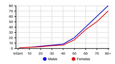
Incidence per 100.000 [gvi]
Symptoms & findings
Symptoms
Anorexia, Arthralgia, Cold skin, Decreased consciousness, Distant heart sounds, Dizziness, Dysphoria, Dyspnea, Edema, Fatigue, Fever, Hepatomegaly, Hypotension, Jugular vein distention, Kussmaul's sign, Myalgia, Night sweats, Pallor, Palpitations, Tachycardia, Tachypnea, Weight loss
Clinical findings
Elevated Jugular Venous Pressure, Friction rub, Inverted T wave, Low voltage QRS, Pericardial effusion, Pleural effusion, PR depression, Pulsus alternans, Pulsus paradoxus, ST elevation
Anamneses
Localized findings
Approach
Blood test (creatine kinase, troponin, coagulation panel, ANA, RF)
Blood gas
ECG
Chest x-ray
Echocardiography
CT thorax
Pericardiocentesis
Treatment
Oxygen [gyy]
Volume expansion with blood, plasma
Bed rest with leg elevation --> increase venous return
Hemodynamically stable patients --> Percutaneous Pericardiocentesis
Hemodynamically unstable patients --> Open Pericardiotomy
Differential diagnoses
Cancer, Cardiomyopathy, Endocarditis, Endomyocardial fibrosis, Heart failure, Myocardial infarction, Myocarditis, Pericarditis, Pleural effusion, Pneumothorax, Pulmonary embolism, Renal failure, Rheumatic fever, Shock, Tension pneumopericardium, Tuberculosis
References
[1] http://emedicine.medscape.com/article/152083 (2014-01-21); [Medscape]
[2] Stashko E, Meer JM. Cardiac Tamponade. [Updated 2023 Aug 7]. In: StatPearls [Internet]. Treasure Island (FL): StatPearls Publishing; 2025 Jan-. Available from: https://www.ncbi.nlm.nih.gov/books/NBK431090/
[3] Al-Ogaili et al. Cardiac Tamponade Incidence, Demographics and in-hospital Outcomes: Analysis of the National Inpatient Sample Database. J Am Coll Cardiol. 2018 Mar, 71 (11_Supplement) A1155.
[4] Ariyarajah V, Spodick DH. Cardiac tamponade revisited: a postmortem look at a cautionary case. Tex Heart Inst J. 2007;34(3):347-51