Diagnosis
Stroke
Sudden death of neurons due to inadequate blood flow.
Also known as: Apoplexia cerebri, Apoplexy, Cerebral infarction, Ischemic stroke
Etiology
Cause [mpt][rfd]
Hemorrhage (10%)
Subarachnoid Hemorrhage (5%): spontaneous (85%), traumatic
Thromboembolism --> Vascular occlusion (85%)
Large-artery stroke: Embolization of atherosclerotic debris originating from the carotid arteries or the heart
Small-vessel lacunar stroke - 15%: Microatheroma or microemboli cause occlusion of the parenchymal branches
Cardioembolic infarction - 20%: Atrial fibrillation, myocardial infarction, mitral stenosis, endocarditis, prosthetic valve, atrial myxoma
Pathophysiology [mpt][rfd]
Embolus pathway: Heart -> Carotid Artery and Vertebral Artery -> Circle of Willis:
Anterior Cerebral Artery (medial frontal lobe)
Contralateral hemiparesis in the lower extremities
Middle Cerebral Artery (lateral frontal lobe, parietal lobe, temporal lobe)
Contralateral hemiparesis in the face and upper extremities
Facial paralysis
Gaze preference toward the side of the lesion
Dysarthria
Aphasia
Neglect
Visual field loss (contralateral inferior quadrantanopia)
Posterior Cerebral Artery (occipital lobe medial portion of the temporal lobe)
Visual cortex: contralateral homonymous hemianopia, cortical blindness
Thalamus: hypersomnolence, cognitive impairment, ocular findings, hypoesthesia, and ataxia
Larger infarcts --> hemisensory loss and hemiparesis
Anmesia
Vertebral artery and basilar artery
Ataxia, vertigo, headache, vomiting, oropharyngeal dysfunction, visual field deficits, abnormal oculomotor findings
Cerebellar Infarction
Ataxia, nausea, vomiting, headache, dysarthria, and vertigo
Insufficient blood supply --> ischemia/cell hypoxia --> irreversible damage (infarction)
Depletion of ATP --> unable to maintain ionic gradients across the cell membrane --> influx of sodium and calcium ions --> passive inflow of water into the cell --> cytotoxic edema
Ischemia --> breakdown of the blood-brain barrier occurring within 4-6 hours after infarction --> proteins and water flood into the extracellular space --> leading to vasogenic edema --> brain swelling and mass effect that peak at 3-5 days and resolve over the next several weeks with resorption of water and proteins
Hemorrhagic transformation occurs in 5% of ischemic strokes
The infarcted tissue eventually undergoes liquefaction necrosis and is removed by macrophages, with the development of parenchymal volume loss.
Risk factors [mpt]
Age, smoking, diabetes mellitus, hypertension, migraine, transient ischemic attacks, atrial fibrillation, hypercholesterolemia, carotid stenosis, physical inactivity, obesity, hormone replacement, heredity
Hypercoagulable states: antiphospholipid antibodies, protein C deficiency, protein S deficiency, pregnancy, polycythemia, thrombocytosis, thrombocytopenia, leukemia, Sickle cell anemia
Fibromuscular dysplasia
Arterial dissections
Vasoconstriction (cocaine, amphetamines)
Complications [mpt][rfd]
Intracranial hemorrhage after ischemic stroke
Deep vein thrombosis
Pulmonary embolism
Aspiration pneumonia
Recurrent seizures
Cerebral edema --> increased ICP
Hydrocephalus
Epidemiology
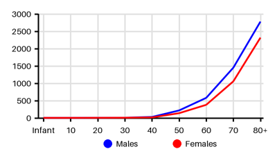
Incidence per 100.000 [2nt][fyp][eiz][d5a][bvq][qf8]
Symptoms & findings
Symptoms
Abducens nerve palsy, Agnosia, Akinesia, Anopsia, Aphasia, Arrhythmia, Ataxia, Blindness, Decreased consciousness, Diplopia, Dysarthria, Dysphagia, Facial nerve paralysis, Fecal incontinence, Headache, Heart murmur, Hyporeflexia, Impotence, Muscle weakness, Nausea, Neck stiffness, Nystagmus, Oculomotor nerve palsy, Paralysis, Paresis, Paresthesia, Rectal sphincter atonia, Scotoma, Seizure, Urinary incontinence, Vascular murmur, Vertigo, Visual disturbances, Vomiting
Clinical findings
Anamneses
Approach
Blood test: Complete blood count, electrolytes, creatinine, coagulation studies, Troponins, PT-INR, toxicology, HCG due to thrombolysis, sedimentation rate, ANA, RF
Blood gas
ECG
CT caput
MR caput
CT angiography
Doppler ultrasonography
Echocardiography
Lumbar puncture
Treatment
The goal of therapy is to preserve areas of reversible tissue damage. [mpt]
Vital signs: [mpt]
Airway
Breathing
Circulation --> Blood pressure <180/105
Labetalol 10 to 20 mg IV; may repeat once
Nicardipine 5 mg/hour IV, increase 2.5 mg/hour (max 15 mg/hour)
Clevidipine 1 to 2 mg/hour IV, double dose every 15 minutes (max 21 mg/hour)
Hydralazine and enalaprilat may be considered
Temperature <38 °C (acetaminophen, exclude infections)
Glucose: insuline to achieve normoglycemia
Nutrition: Early enteric feeding is encouraged (dysphagia -> nasogastric tube)
Thrombosis prophylaxis: heparin or low molecular weight heparin
Ischemic stroke recanalization strategies: Effective if performed quickly
Antiplatelet/anticoagulation:
Aspirin is recommended within 24 to 48 hours of symptom onset
DVT-prophylaxis: low-dose heparin or low molecular weight heparin
Atrial fibrillation: oral anticoagulation within 4 to 14 days
IV alteplase/tenecteplase (within 4.5 hours of stroke onset)
Mechanical thrombectomy (within 6 hours of stroke onset) if large vessel occlusion
Intracerebral Hemorrhage [rfd]
Discontinued anticoagulation immediately, rapid reversal
Platelet transfusion if treated with antiplatelet or indication for neurosurgery
Surgery:
Minimally invasive hematoma evacuation with endoscopic/stereotactic aspiration
External ventricular drain insertion
Craniotomy
Subarachnoid Hemorrhage [rfd]
Early (<72 hours) endovascular coiling (or clipping) of aneurysm to decrease the risk of rebleeding
Seizure --> antiepileptic drugs
Cerebellar/Cerebral edema:
Peaks in 3 to 5 days after an ischemic stroke
Correlate with the size of the stroke
May increase ICP/herniation/hydrocephalus --> apnea, pupillary dilatation, death
Treatment: ventriculostomy, decompressive craniectomy, surgical evacuation
Post-stroke seizure: Antiepileptic drugs for 1 month (or longer if late seizures)
Cardiac evaluation: Treatment of atrial fibrillation
Differential diagnoses
Amphetamine toxicity, Bell's Palsy, Benign paroxysmal positional vertigo, Brain abscess, Brain tumor, Cerebral venous sinus thrombosis, Cocaine toxicity, Drug side effects, Epilepsy, Hepatic encephalopathy, Hyperglycemic hyperosmolar nonketotic state, Hypoglycemia, Hyponatremia, Intracranial hemorrhage, Migraine, Multiple sclerosis, Myocardial infarction, Postseizure encephalopathy, Psychotropic drugs, Sepsis, Subarachnoid hemorrhage, Syncope, Transient global amnesia, Transient ischemic attack, Wernicke encephalopathy
References
[1] Lui F, Hui C, Khan Suheb MZ, et al. Ischemic Stroke. Updated 2025 Feb 21: https://www.ncbi.nlm.nih.gov/books/NBK499997/
[2] Tadi P, Lui F. Acute Stroke. [Updated 2023 Aug 17]. In: StatPearls [Internet]. Treasure Island (FL): StatPearls Publishing; 2025 Jan-. Available from: https://www.ncbi.nlm.nih.gov/books/NBK535369/
[3] Nawaz B, Eide GE, Fromm A, Øygarden H, Sand KM, Thomassen L, Næss H, Waje-Andreassen U. Young ischaemic stroke incidence and demographic characteristics - The Norwegian stroke in the young study - A three-generation research program. Eur Stroke J. 2019 Dec;4(4):347-354.
[4] Kissela BM, Khoury JC, Alwell K, Moomaw CJ, Woo D, Adeoye O, Flaherty ML, Khatri P, Ferioli S, De Los Rios La Rosa F, Broderick JP, Kleindorfer DO. Age at stroke: temporal trends in stroke incidence in a large, biracial population. Neurology. 2012 Oct 23;79(17):1781-7.
[5] Ramirez L, Kim-Tenser MA, Sanossian N, Cen S, Wen G, He S, Mack WJ, Towfighi A. Trends in Acute Ischemic Stroke Hospitalizations in the United States. J Am Heart Assoc. 2016 May 11;5(5):e003233.
[6] Hong KS, Bang OY, Kang DW, Yu KH, Bae HJ, Lee JS, Heo JH, Kwon SU, Oh CW, Lee BC, Kim JS, Yoon BW. Stroke statistics in Korea: part I. Epidemiology and risk factors: a report from the korean stroke society and clinical research center for stroke. J Stroke. 2013 Jan;15(1):2-20.
[7] Feigin VL, Krishnamurthi R, Nair B, Rautalin I, Parag V, Anderson CS, Arroll B, Barber PA, Barker-Collo S, Bennett D, Brown P, Cadilhac DA, Douwes J, Exeter D, Ranta A, Ratnasabapathy Y, Swain A, Tautolo ES, Te Ao B, Thrift A, Tunnage B. Trends in stroke incidence, death, and disability outcomes in a multi-ethnic population: Auckland regional community stroke studies (1981-2022). Lancet Reg Health West Pac. 2025 Mar 10;56:101508.
[8] Chau PH, Woo J, Goggins WB, Tse YK, Chan KC, Lo SV, Ho SC. Trends in stroke incidence in Hong Kong differ by stroke subtype. Cerebrovasc Dis. 2011;31(2):138-46.
[9] http://emedicine.medscape.com/article/1916852 (2014-02-05); [Medscape]