Diagnosis
Gallbladder cancer
Malignant neoplasm of the gallbladder.
Etiology
Cause/Pathophysiology [rtu]
Chronic inflammation (or toxins and direct injury) --> DNA mutations (K-ras, TP53, CDKN2a, c-erb-b2)
Cholesterol gallstones (most common)
Primary sclerosing cholangitis
Ulcerative colitis
Crohns disease
Liver parasite
Chronic Salmonella typhi/paratyphi infection
Helicobacter infection
Extrinsic carcinogens:
Medications: methyldopa, isoniazid
Work exposure: methylcellulose, radon
Lifestyle: cigarette smoking, obesity, high carbohydrate intake
Polyps --> dysplasia --> invasive gallbladder cancer (usually adenocarcinoma)
Other rare types: squamous, adenosquamous, and neuroendocrine tumors
Grim prognosis with 5 year survival rate 38% [m49] due to: [rtu]
Aggressive tumor biology
Complex anatomical location
Advanced stage at diagnosis
Risk factors [rtu]
Woman
Gallstones
Porcelain gallbladder (gallbladder calcification)
Choledochal cysts
Gallbladder polyps
Occupational chemical exposure
Estrogens
Anomalous pancreatobiliary duct junction
Hereditary syndromes with an increased incidence of gallbladder cancer: Gardner syndrome, neurofibromatosis type I, and hereditary nonpolyposis colon cancer.
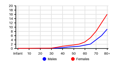
Epidemiology
Incidence per 100.000 [fq5]
Symptoms & findings
Symptoms
Anorexia, Courvoisier's sign, Fever, Hepatomegaly, Icterus, Lymphadenopathy, Nausea, Sister Mary Joseph's nodule, Virchow's node, Vomiting, Weight loss
Clinical findings
Anemia, Cholestasis, Decreased Albumin, Elevated ALP, Elevated Bilirubin, Elevated CA 19-9, Elevated CEA, Elevated GGT, Elevated PT-INR, Jaundice
Anamneses
None listed.
Localized findings
Approach
Blood test: CA 19-9 (elevated), CEA (not specific for gallbladder cancer), liver function tests
Ultrasound abdomen
CT abdomen
MR abdomen
MRCP
ERCP
PTC
Classification [rtu]
T1: Tumor invades lamina propria or muscular layer
T1a: Tumor invades lamina propria
T1b: Tumor invades muscle layer
T2: Tumor invades the perimuscular connective tissue
T2a: Tumor invades the perimuscular connective tissue on the peritoneal side, without the involvement of the serosa (visceral peritoneum)
T2b: Tumor invades the perimuscular connective tissue on the hepatic side, with no extension into the liver
T3: Tumor perforates the serosa (visceral peritoneum) and/or directly invades the liver and/or 1 other adjacent organ or structure, such as the stomach, duodenum, colon, pancreas, omentum, or extrahepatic bile ducts
T4: Tumor invades main portal vein or hepatic artery or invades 2 or more extrahepatic organs or structures
N (regional lymph nodes)
NX: Regional lymph nodes cannot be assessed
N0: No regional lymph node metastasis
N1: Metastases to 1 to 3 regional lymph nodes
N2: Metastases to ≥4 or more regional lymph nodes
M (distant metastasis)
M0: No distant metastasis
M1: Distant metastasis [3]
Treatment
Early-stage disease (a minority of patients): [rtu][m49]
T1a tumors (incidentally detected): Tumors confined to the mucosa require only a cholecystectomy
T1b, T2, and T3 tumors: En-bloc resection of the gallbladder including resection of the adjacent segments 4b and 5 with adequate surgical margins + portal lymphadenectomy + adjuvant radiotherapy to control microscopic residual deposits of carcinoma in the tumor bed and regional lymph nodes
Extended hepatic (beyond segments 4b and 5) and biliary resections increase morbidity and mortality without any survival benefit
Neoadjuvant chemotherapy: Some patients may have an adequate response that allows for subsequent resection
Unresectable disease due to locally advanced and metastatic disease:
Palliation with radiation therapy and/or chemotherapy
Differential diagnoses
Acalculous cholecystitis, Ampullary carcinoma, Benign bile duct tumor, Bile duct stricture, Biliary obstruction, Carcinoma of the ampulla of Vater, Cholangiocarcinoma, Cholangitis, Cholecystitis, Choledochal cysts, Choledocholithiasis, Cholelithiasis, Gallbladder mucocele, Gallbladder volvulus, Gastrinoma, Glucagonoma, Hepatocellular carcinoma, Insulinoma, Liver abscess, Pancreatic cancer, Primary biliary cirrhosis, Primary sclerosing cholangitis, VIPoma
References
[1] Menon G, Babiker HM. Gallbladder Carcinoma. [Updated 2024 Aug 17]. In: StatPearls [Internet]. Treasure Island (FL): StatPearls Publishing; 2025 Jan-. Available from: https://www.ncbi.nlm.nih.gov/books/NBK442002/
[2] http://emedicine.medscape.com/article/278641 (2014-01-02); [Medscape]
[3] https://www.cancerresearchuk.org/health-professional/cancer-statistics/statistics-by-cancer-type/gallbladder-cancer/incidence#heading-One
[4] [uptodate.com]
[5] Dulskas A, Cerkauskaite D, Patasius A, Smailyte G. Incidence, Mortality, and Survival Trends in Cancer of the Gallbladder and Extrahepatic Bile Ducts in Lithuania. Medicina (Kaunas). 2023 Mar 27;59(4):660.