Diagnosis
Multiple myeloma
A malignant proliferation of plasma cells involving more than 10% of the bone marrow.
Etiology
Cause [peb]
Unknown
Spontaneous mutation in the promoter genes or oncogenes NRAS, KRAS, and BRAF
Pathophysiology [peb]
Monoclonal gammopathy of undetermined significance (MGUS):
Monoclonal immunoglobulins in the blood or urine without end-organ damage
Plasma cell proliferation --> overproduction of monoclonal IgG, IgA, light chains
Risk of progression to multiple myeloma
MGUS --> Multiple myeloma:
Genetic alterations --> increased expression of promoter genes --> resistance to apoptosis --> higher plasma cell proliferation and population >10% of the bone marrow
Myeloma cells --> activation of osteoclasts and suppression of osteoblasts
Osteolytic lesions in the vertebrae, ribs, skull, pelvis, femur, clavicle and scapula
Risk factors [peb]
Obesity
Alcohol
Insecticides, organic solvents, agent orange
Radiation exposure
Complications [peb]
Renal failure
Hyperviscosity (stroke, myocardial infartcion, venous thromboembolism)
Pancytopenia: Anemia, thrombocytopenia, and leukopenia
Neuropathy
Infection
Skeletal lesions: pathological fractures, spinal cord compression
Hypercalcemia
Epidemiology
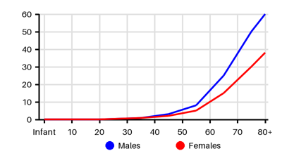
Incidence per 100.000 [eia][2h5][6aa]
Symptoms & findings
Symptoms
Ataxia, Epistaxis, Fasciculations, Headache, Hepatomegaly, Muscle weakness, Paresis, Paresthesia, Somnolence, Splenomegaly, Weakness, Weight loss
Clinical findings
Anemia, Bence Jones proteinuria, Elevated Creatinine, Elevated CRP, Elevated Sedimentation Rate, Elevated Uric Acid, Hypercalcemia, Hypergammaglobulinemia, Hyperproteinemia, Lytic bone lesion, Macroglossia, M-component, Pathologic fracture, Rouleaux formation, Thrombocytopenia
Anamneses
None listed.
Localized findings
Approach
Blood test: Hb, WBC, Trc, SR, CRP, BUN, creatinine, uric acid, calcium, LDH, beta-2 microglobulin
Urine:
24-hour proteinuria
Urine protein electrophoresis, Bence Jones protein (lambda light chains)
Urine immunofixation electrophoresis
Serum protein electrophoresis:
Serum immunoglobulins
Serum protein electrophoresis
Serum immunofixation electrophoresis
Serum-free light chain assay
Blood smear
Bone marrow aspirate and biopsy:
Immunohistochemistry and/or flow cytometry,
Cytogenetics
Plasma cell FISH
Bone biopsy
X-ray
MRI
Whole body PET-CT
Diagnostic criteria (International Myeloma Working Group 2014) [peb]
Clonal bone marrow plasma cells > 10% on bone marrow biopsy
Or if a biopsy-proven plasmacytoma
And one of the following criteria for end-organ damage:
Serum calcium level >2.75 mmol/L (>11 mg/dL)
Renal insufficiency (creatinine > 2 mg/dL [> 177 umol/L] or clearance < 40 mL/min)
Anemia (Hb <10 g/dL)
One or more osteolytic bone lesions
Bone marrow plasma cells equal to 60%
Involved/uninvolved serum free light chain ratio ≥ 100
Abnormal MRI with more than one focal lesion >5 mm
Treatment
Treatment is dependant on subtype: [q5a][peb]
Monoclonal gammopathy of undetermined significance (MGUS): 2-3% of the elderly
Premalignant state
Bone marrow plasma cell concentration < 10%
No end-organ damage
Active surveillance or watchful waiting
Solitary plasmacytoma (lesion made up of clonal plasma cells)
Normal bone marrow
No end-organ damage
Treatment: Radiation therapy or surgery if structural instability
Smoldering multiple myeloma (SMM)
Monoclonal protein is greater than or equal to 3 g/dl
Clonal bone marrow plasma cells between 10% to 59%
No end-organ damage
Treatment: Active surveillance or watchful waiting
Symptomatic MM
Supportive treatment: Hypercalcemia, renal dysfunction, spinal cord compression
Young patients --> Transplantation
Elderly patients --> Triplet regime:
lenalidomide/dexamethasone/Bortezomib or Carfilzomib
Bortezomib/cyclophosphamide/dexamethasone (if kidney failure)
Carfilzomib/cyclophosphamide/dexamethasone (peripheral neuropathy)
Differential diagnoses
Amyloidosis, Breast cancer, Leukemia, Lung cancer, Lymphoma, Macroglobulinemia, Metastatic bone tumors, Polyneuropathy, Prostate cancer, Renal cell carcinoma, Renal failure, Thyroid cancer
References
[1] Albagoush SA, Shumway C, Azevedo AM. Multiple Myeloma. [Updated 2023 Jan 30]. In: StatPearls [Internet]. Treasure Island (FL): StatPearls Publishing; 2025 Jan-. Available from: https://www.ncbi.nlm.nih.gov/books/NBK534764/
[2] Ludwig H, Novis Durie S, Meckl A, Hinke A, Durie B. Multiple Myeloma Incidence and Mortality Around the Globe; Interrelations Between Health Access and Quality, Economic Resources, and Patient Empowerment. Oncologist. 2020 Sep;25(9):e1406-e1413
[3] Turesson I, Velez R, Kristinsson SY, Landgren O. Patterns of multiple myeloma during the past 5 decades: stable incidence rates for all age groups in the population but rapidly changing age distribution in the clinic. Mayo Clin Proc. 2010 Mar;85(3):225-30
[4] Mousavi, S.E., Ilaghi, M., Aslani, A. et al. A population-based study on incidence trends of myeloma in the United States over 2000–2020. Sci Rep 13, 20705 (2023)
[5] http://emedicine.medscape.com/article/204369 (2014-01-02); [Medscape]