Diagnosis
Subarachnoid hemorrhage
A life-threatening brain hemorrhage with accumulation of blood between the arachnoid and pia mater.
Etiology
Cause [za1]
Traumatic
Non-traumatic:
Rupture of aneurysm (85%)
AV-malformation (10%)
Intracranial arterial dissection
Other causes: Cocaine abuse, sickle cell anemia, anticoagulant therapy, bleeding disorders, pituitary apoplexy, cerebral amyloid angiopathy, cerebral venous thrombosis, moyamoya disease, cerebral vasculitis
Pathophysiology [za1]
Subarachnoid space:
Between the arachnoid membrane and the pia mater
Consists of the cerebrospinal fluid and the cerebral blood vessels
Hemodynamic stress --> inflammation --> formation of intracranial aneurysm
Arterial junctions
Bifurcations
Abrupt vascular angles
Large aneurysms may compress the adjacent structures --> neurological signs
Law of Laplace: wall stress = (Pressure x Radius) / (2 x wall thickness)
Rupture:
Elevated intracranial pressure --> reduced cerebral perfusion --> cerebral ischemia
Mechanical blockage of the CSF flow --> hydrocephalus
Cerebral vasospasm --> delayed cerebral ischemia
Risk factors [za1]
Large aneurysm (>5 mm)
Older age (>60 years)
Atherosclerosis
Hypertension
Cigarette smoking
Family history
Alcohol
Sympathomimetic drugs
Estrogen deficiency
Polycystic kidney disease
Complications [za1]
Vasospasm (40-60%)
Hydrocephalus (30%)
Rebleeding (9-17%)
Seizure
Increased intracranial pressure
Brain herniation
Cerebral infarction
Neurogenic pulmonary edema
Death
Epidemiology
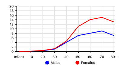
Incidence per 100.000 [w5s][tmd][7sd][cns][8od][za1]
Symptoms & findings
Symptoms
Anisocoria, Anorexia, Arrhythmia, Bradycardia, Cardiac arrest, Cheyne-Stokes respiration, Coma, Confusion, Cushing's triad, Decreased consciousness, Deviated gaze, Diplopia, Dysphasia, Headache, Hypertension, Intraocular hemorrhage, Irregular respiration, Myalgia, Nausea, Neck stiffness, Paralysis, Paresis, Paresthesia, Photophobia, Ptosis, Seizure, Unconsciousness, Vascular murmur, Vomiting, Wide pulse pressure
Clinical findings
Decreased Blood Urea Nitrogen, Decreased Uric Acid, Elevated Intracranial Pressure, Hyponatremia, Hyposmolality, Subarachnoid blood
Anamneses
Localized findings
Approach
Blood test
Blood gas
Ophtalmoscopy
CT angiography
MR angiography
Cerebral angiography: detecting vasospasm
Transcranial Doppler: monitoring vasospasm
Lumbar puncture
Xanthochromia
EEG: subclinical seizures or nonconvulsive status epilepticus
ECG
Treatment
Goal of treatment: prevent rebleeding, hydrocephalus and vasospasm
Initial treatment: [za1]
Airway: Decreased consciousness --> Intubation
Breathing: Respiratory failure --> Oxygen or mechanical ventilation
Circulation: Blood pressure <160 mm Hg (prevention of rebleeding/vasospasm):
Labetalol 5-20 mg IV every 15 minutes
Hydralazine 20-40 mg every 30 to 60 minutes
Nicardipine 5-15 mg/hr continuous IV infusion
Clevidipine 1-2 mg/hr and repeated every 90 seconds
Nimodipine (oral) 60 mg every 4 hours
Supportive treatment: [za1]
Osmotic diuretics (Mannitol)
Antiepileptics
Antibiotics
Anticoagulants: discontinue all anti-thrombotic agents and reverse all anticoagulation until the aneurysm is definitively repaired
Pain control: Acetaminophen 1000 mg IV, opioid 2-4 mg IV every 1 to 2 hours
Interventional options: endovascular coiling
Surgery:
Clipping of the aneurysm
Intraventricular external drainage device to reduce the ICP/hydrocephalus
Differential diagnoses
Arteriovenous malformation, Brain tumor, Cerebral aneurysm, Epidural hematoma, Hydrocephalus, Intracerebral hemorrhage, Meningitis, Multiple sclerosis, Sarcoidosis, Stroke, Subdural hematoma, Syndrome of inappropriate antidiuretic hormone secretion, Toxoplasmosis
References
[1] Ziu E, Khan Suheb MZ, Mesfin FB. Subarachnoid Hemorrhage. Updated 2023 Jun 1: https://www.ncbi.nlm.nih.gov/books/NBK441958/
[2] Linn FH, Rinkel GJ, Algra A, van Gijn J. Incidence of subarachnoid hemorrhage: role of region, year, and rate of computed tomography: a meta-analysis. Stroke. 1996 Apr;27(4):625-9.
[3] Øie LR, Solheim O, Majewska P, Nordseth T, Müller TB, Carlsen SM, Jensberg H, Salvesen Ø, Gulati S. Incidence and case fatality of aneurysmal subarachnoid hemorrhage admitted to hospital between 2008 and 2014 in Norway. Acta Neurochir (Wien). 2020 Sep;162(9):2251-2259.
[4] Sun T, Chen S, Wu K, Sun M, Zhang X, You C. Trends in Incidence and Mortality of Stroke in China From 1990 to 2019. Front Neurol. 2021 Nov 22;12:759221.
[5] Lv B, Lan JX, Si YF, Ren YF, Li MY, Guo FF, Tang G, Bian Y, Wang XH, Zhang RJ, Du ZH, Liu XF, Yu SY, Tian CL, Cao XY, Wang J. Epidemiological trends of subarachnoid hemorrhage at global, regional, and national level: a trend analysis study from 1990 to 2021. Mil Med Res. 2024 Jul 11;11(1):46.
[6] Xia C, Hoffman H, Anikpezie N, Philip K, Wee C, Choudhry R, Albright KC, Masoud H, Beutler T, Schmidt E, Gould G, Patel SD, Akano EO, Morris N, Chaturvedi S, Aneni E, Lamikanra O, Chin L, Latorre JG, Otite FO. Trends in the Incidence of Spontaneous Subarachnoid Hemorrhages in the United States, 2007-2017. Neurology. 2023 Jan 10;100(2):e123-e132.