Diagnosis
Hypovolemia
Abnormally decreased intravascular volume, leading to systemic hypoperfusion, tissue hypoxia, and, if untreated, multiorgan failure.
Also known as: Hypovolemic Shock
Etiology
Cause [iwf][foe]
Renal
Diuretic excess
Mineralocorticoid deficiency
Ketonuria
Osmotic diuresis
Cerebral salt wasting syndrome
Salt-wasting nephropathies
Gastrointestinal
Vomiting
Diarrhea
Bowel obstruction (GI secretes 3-6 liters of fluid daily)
Stoma/fistula
Skin
Burns
Hot and dry climate
Third spaceing
Sepsis
Pancreatitis
Trauma
Bleeding (internal hemorrhage, GI bleeding, uterine bleeding)
Postoperative phase
Anaphylaxis
Pathophysiology [foe]
Blood pressure = Cardiac output × systemic vascular resistance
Cardiac output = heart rate x stroke volume
Classification
Mild hypovolemia: 3% to 5% volume loss
Moderate hypovolemia: 6% to 9% volume loss
Severe hypovolemia: >10% volume loss
Hypovolemia: reduced SV --> reduction of CO:
Tissue hypoxia
Metabolic acidosis
Anaerobic metabolism
Elevated lactate levels
Progressive cellular damage
Compensation:
Increased SVR (vasoconstriction and redistribution to the brain, heart, and kidneys)
Increased heart rate and myocardial contractility
Pituitary gland: Antidiuretic hormone --> promotes salt and water resorption
Renin --> angiotensinogen --> angiotensin I --> angiotensin II:
Vasoconstrictor
Stimulate aldosterone --> sodium reabsorption and water conservation
Complications [iwf]
Shock
Ischemic stroke
Myocardial infarction
Stroke
Sheehan syndrome
Liver failure
Acute Renal failure
Multi-organ failure
Death
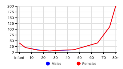
Epidemiology
Incidence per 100.000 [zqv]
Symptoms & findings
Symptoms
Anuria, Burns, Cyanosis, Dehydration, Diarrhea, Dizziness, Dry mucous membranes, Fatigue, Flat neck veins, Headache, Hypotension, Hypothermia, Lethargy, Muscle cramps, Muscle weakness, Narrow pulse pressure, Oliguria, Poor skin turgor, Prolonged capillary refill, Somnolence, Tachycardia, Thirst, Vomiting, Weakness, Weight loss
Clinical findings
Elevated Hematocrit, Elevated Hemoglobin, Polycythemia
Anamneses
None listed.
Approach
Blood test: Hemoglobuline, hematocrit, creatinine, blood urea nitrogen, sodium (Na), potassium (K), glucose
Urine: Sodium (Na), volume, osmolality
Blood gas: Lactate, pH
Weight
Leg raise
Ultrasonography: IVC or jugular vein diameter and collapsibility
CVP
Volumetric preload parameter: Intrathoracic blood volume index
Treatment
Fluid resuscitation [iwf] [foe]
Chronic hypovolemia --> gradual resuscitation
Excessive fluid resuscitation --> risk of cardiac and pulmonary complications
Warm isotonic crystalloid solution of 30 ml/kg infused rapidly
If hemorrhagic shock --> Transfusion 1:1:1 of plasma to platelets to RBC
Effective resuscitation can be monitored by heart rate, blood pressure, urine output, mental status, and peripheral edema
Vasopressor after sufficient volume resuscitation
Urinary catheter to measure urine output
Arterial line to measure arterial blood pressure
Differential diagnoses
Abdominal compartment syndrome, Acute liver failure, Acute pancreatitis, Amniotic fluid embolism, Anaphylaxis, Anemia, Aortic regurgitation, Aortic stenosis, Aortoenteric fistula, Arrhythmia, Atrial myxoma, Beriberi, Burn wound, Cardiac contusion, Cardiac tamponade, Cardiomyopathy, Diarrhea, Drug side effects, Fat embolism syndrome, Gastroenteritis, Gastrointestinal hemorrhage, Heart failure, Hemothorax, Hypoaldosteronism, Hypoglycemia, Internal hemorrhage, Myocardial infarction, Myocarditis, Neurogenic shock, Perforated ulcer, Pericarditis, Pneumothorax, Pregnancy, Pulmonary embolism, Pulmonary hypertension, Renal failure, Retroperitoneal hemorrhage, Ruptured aortic aneurysm, Ruptured ectopic pregnancy, Sepsis, Shock, Takotsubo cardiomyopathy, Tension penumothorax, Thyrotoxicosis, Toxic shock syndrome, Trauma
References
[1] Melendez Rivera JG, Anjum F. Hypovolemia: https://www.ncbi.nlm.nih.gov/books/NBK565845/
[2] Taghavi S, Nassar Ak, Askari R. Hypovolemic Shock: https://www.ncbi.nlm.nih.gov/books/NBK513297/
[3] Gitz Holler J, Jensen HK, Henriksen DP, Rasmussen LM, Mikkelsen S, Pedersen C, Lassen AT. Etiology of Shock in the Emergency Department: A 12-Year Population-Based Cohort Study. Shock. 2019 Jan;51(1):60-67.